A case of cerebral tuberculoma mimicking neurocysticercosis
- PMID: 29123884
- PMCID: PMC5674469
- DOI: 10.1002/ams2.272
A case of cerebral tuberculoma mimicking neurocysticercosis
Abstract
Case: A 42-year-old Peruvian woman residing in Japan for 11 years with a family history of neurocysticercosis presented to our intensive care unit with fever and intense headache.Computed tomography indicated multiple micronodular lesions in the brain parenchyma, and cerebral tuberculoma and neurocysticercosis were considered in the differential diagnosis. Neurocysticercosis was initially suspected, and oral praziquantel was initiated. However, because of a high adenosine deaminase level in the cerebrospinal fluid and positive peripheral blood interferon gamma release test result, cerebral tuberculoma was subsequently considered.
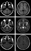
Outcome: Antituberculous drugs with steroids were initiated on day 10, after which the symptoms gradually resolved; the patient was discharged on day 29. Gadolinium-contrast magnetic resonance imaging 8 months later showed reduced nodular shadows, confirming cerebral tuberculoma.
Conclusion: Immediate diagnosis and treatment are imperative for cerebral tuberculoma, a lethal infection. Considering the recent increases in immigration worldwide, increased cases of tuberculoma mimicking neurocysticercosis are expected.
Keywords: Adenosine deaminase; immigrants; interferon gamma release tests; intracranial tuberculoma; neurocysticercosis.
Figures


References
-
- Gothi R. Consider tuberculoma and cysticercosis in the differential diagnosis of brain tumour in tropical countries. BMJ 2013; 347: f6604. - PubMed
-
- Oncul O, Baylan O, Mutlu H, Cavuslu S, Doganci L. Tuberculous meningitis with multiple intracranial tuberculomas mimicking neurocysticercosis clinical and radiological findings. Jpn. J. Infect. Dis. 2005; 58: 387–9. - PubMed
-
- Mukherjee S, Das R, Begum S. Tuberculoma of the brain‐A diagnostic dilemma: magnetic resonance spectroscopy a new ray of hope. J. Assoc. Chest Physicians 2015; 3: 3–8.
-
- Kim SH, Cho OH, Park SJ et al Rapid diagnosis of tuberculous meningitis by T cell‐based assays on peripheral blood and cerebrospinal fluid mononuclear cells. Clin. Infect. Dis. 2010; 50: 1349–58. - PubMed
Publication types
LinkOut - more resources
Full Text Sources
Other Literature Sources
Research Materials

