

T2 mapping and post-contrast T1 (dGEMRIC) of the patellar cartilage: 12-year follow-up after patellar stabilizing surgery in childhood
- PMID: 29123919
- PMCID: PMC5661686
- DOI: 10.1177/2058460117738808
T2 mapping and post-contrast T1 (dGEMRIC) of the patellar cartilage: 12-year follow-up after patellar stabilizing surgery in childhood
Abstract
Background: Cartilage degeneration has been reported after recurrent patellar dislocation. However, effects of surgical stabilization in childhood have not yet been described.
Purpose: To examine the cartilage quality in very young adults operated with a patellar stabilizing procedure due to recurrent patellar dislocation in childhood, and evaluate if cartilage quality correlates with clinical parameters and patient-reported outcomes.
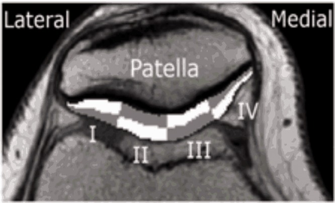
Material and methods: Seventeen patients were investigated ≥ 5 years (mean = 11.6 years) after patellar stabilizing surgery in childhood. Pre-contrast T2 relaxation times were analyzed in four superficial and four deep patellar cartilage regions of both knees. Two hours after 0.2 mM/kg Gd-DTPA2 i.v., post-contrast T1 (T1(Gd)) was analyzed in the same regions. Patient-reported outcomes (KOOS, Kujala, and Tegner scores) and recurrence rates were evaluated.
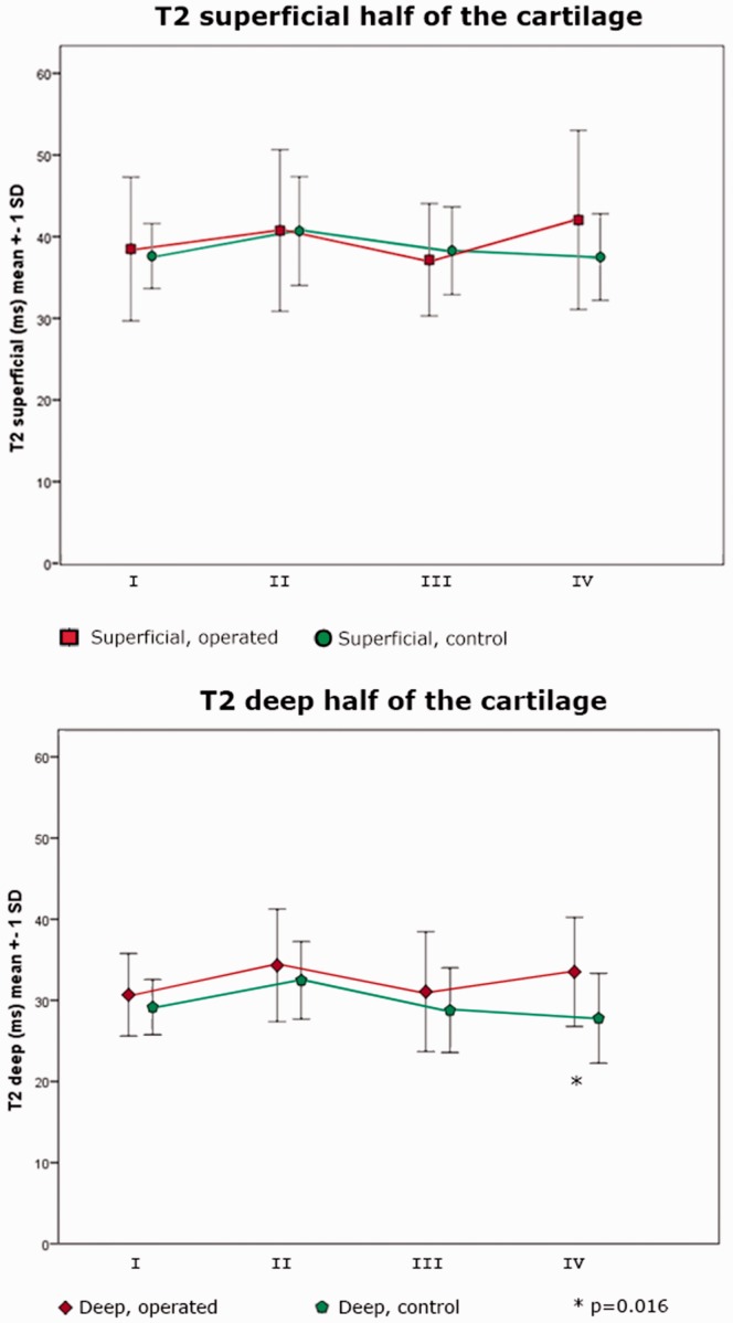
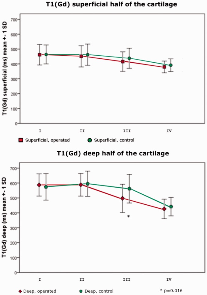
Results: Comparing operated to healthy side, neither T2 nor dGEMRIC differed between the operated and the reference knee regarding the superficial half of the cartilage. In the deep half of the cartilage, T1(Gd) was shorter in the central part of the cartilage, whereas T2 was longer medially (P < 0.05). A low score in the KOOS subscales Symptom and Sports & Recreation, was correlated to the degenerative changes detected by T1(Gd) (r = 0.5, P = 0.041).
Conclusion: In general, our findings demonstrate good cartilage quality 12 years after patellar stabilizing surgery during childhood. The subtle changes in T2 and T1(Gd) in the deep cartilage layer may be a result of altered biomechanics, although very early degenerative changes cannot be excluded. The short T1(Gd) centrally may reflect lower glycosaminoglycan content, whereas the increase in T2 medially indicates increased cartilage hydration.
Keywords: T2; dGEMRIC; patellar dislocation; post-traumatic osteoarthritis.
Figures
Similar articles
-
Pre- and postcontrast T1 and T2 mapping of patellar cartilage in young adults with recurrent patellar dislocation.Magn Reson Med. 2015 Nov;74(5):1363-9. doi: 10.1002/mrm.25511. Epub 2014 Nov 24. Magn Reson Med. 2015. PMID: 25421491
-
In vivo biochemical 7.0 Tesla magnetic resonance: preliminary results of dGEMRIC, zonal T2, and T2* mapping of articular cartilage.Invest Radiol. 2008 Sep;43(9):619-26. doi: 10.1097/RLI.0b013e31817e9122. Invest Radiol. 2008. PMID: 18708855
-
Degeneration of patellar cartilage in patients with recurrent patellar dislocation following conservative treatment: evaluation with delayed gadolinium-enhanced magnetic resonance imaging of cartilage.Osteoarthritis Cartilage. 2009 Dec;17(12):1546-53. doi: 10.1016/j.joca.2009.05.001. Epub 2009 May 18. Osteoarthritis Cartilage. 2009. PMID: 19481191
-
Temporal in vivo assessment of fresh osteochondral allograft transplants to the distal aspect of the femur by dGEMRIC (delayed gadolinium-enhanced MRI of cartilage) and zonal T2 mapping MRI.J Bone Joint Surg Am. 2014 Apr 2;96(7):564-72. doi: 10.2106/JBJS.K.01456. J Bone Joint Surg Am. 2014. PMID: 24695923 Clinical Trial.
-
Use of quantitative MRI for the detection of progressive cartilage degeneration in a mini-pig model of osteoarthritis caused by anterior cruciate ligament transection.J Magn Reson Imaging. 2015 Oct;42(4):1032-8. doi: 10.1002/jmri.24862. Epub 2015 Feb 6. J Magn Reson Imaging. 2015. PMID: 25656460
Cited by
-
Translation and Validation of the Spanish Version of the Banff Patellofemoral Instability Instrument 2.0.Orthop J Sports Med. 2024 Dec 13;12(12):23259671241298306. doi: 10.1177/23259671241298306. eCollection 2024 Dec. Orthop J Sports Med. 2024. PMID: 39678439 Free PMC article.
-
Adaptation of the Banff Patellofemoral Instability Instrument (BPII) 2.0 into Swedish.Acta Orthop. 2023 Oct 31;94:537-542. doi: 10.2340/17453674.2023.21194. Acta Orthop. 2023. PMID: 37905565 Free PMC article.
-
Elevated Patellofemoral and Tibiofemoral T1ρ Relaxation Times Following a First Time Patellar Dislocation.Cartilage. 2022 Apr-Jun;13(2):19476035221102570. doi: 10.1177/19476035221102570. Cartilage. 2022. PMID: 35676874 Free PMC article.
-
MRI T2 and T1ρ relaxation in patients at risk for knee osteoarthritis: a systematic review and meta-analysis.BMC Musculoskelet Disord. 2019 May 1;20(1):182. doi: 10.1186/s12891-019-2547-7. BMC Musculoskelet Disord. 2019. PMID: 31039785 Free PMC article.
References
-
- Askenberger M, Ekström W, Finnbogason T, et al. Occult intra-articular knee injuries in children with hemarthrosis. Am J Sports Med 2014; 42: 1600–1606. - PubMed
-
- Lewallen L, Mcintosh A, Dahm D. First-time patellofemoral dislocation: risk factors for recurrent instability. J Knee Surg 2014; 1: 1–7. - PubMed
-
- Watanabe A, Obata T, Ikehira H, et al. Degeneration of patellar cartilage in patients with recurrent patellar dislocation following conservative treatment: evaluation with delayed gadolinium-enhanced magnetic resonance imaging of cartilage. Osteoarthr Cartil 2009; 17: 1546–1553. - PubMed
-
- Bengtsson Moström E, Lammentausta E, Finnbogason T, et al. Pre- and postcontrast T1 and T2 mapping of patellar cartilage in young adults with recurrent patellar dislocation. Magn Reson Med 2015; 74: 1363–1369. - PubMed
-
- Eagle S, Potter HG, Koff MF. Morphologic and quantitative magnetic resonance imaging of knee articular cartilage for the assessment of post-traumatic osteoarthritis. J Orthop Res 2017; 35: 412–423. - PubMed
LinkOut - more resources
Full Text Sources
Other Literature Sources
