

Targeting IL-5Rα with antibody-conjugates reveals a strategy for imaging and therapy for invasive bladder cancer
- PMID: 29123949
- PMCID: PMC5665064
- DOI: 10.1080/2162402X.2017.1331195
Targeting IL-5Rα with antibody-conjugates reveals a strategy for imaging and therapy for invasive bladder cancer
Abstract
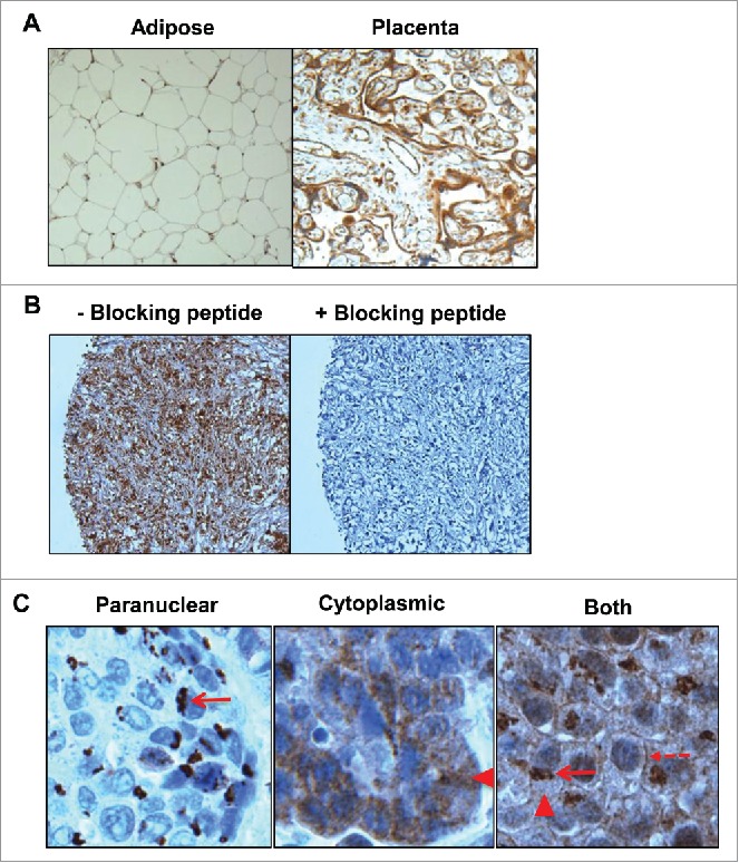
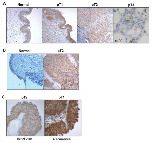
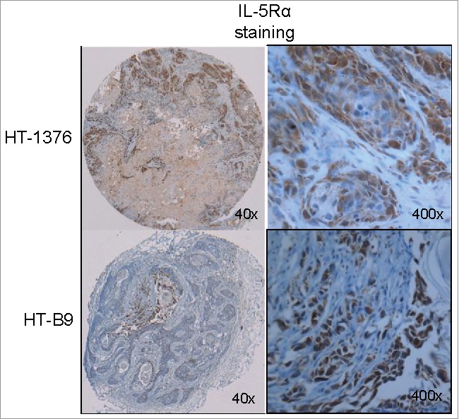
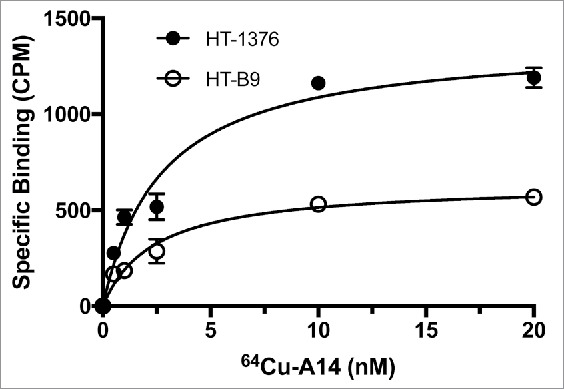
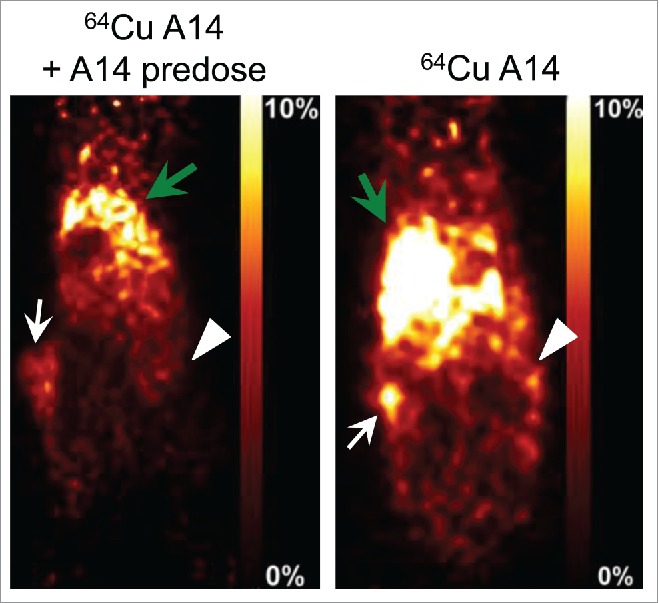
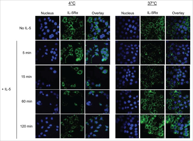
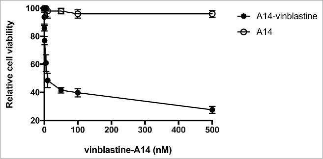
Despite the high interest and concern due to an increasing incidence and death rate, patients who develop muscle invasive bladder cancer (MIBC) have few options available. However, the past decade has produced many candidate bladder tumor-specific markers but further development of these markers is still needed for creating effective targeted medications to solve this urgent need. Interleukin-5 receptor α-subunit (IL-5Rα) has recently been reported to be involved in MIBC progression. Thus, we aimed to validate IL-5Rα as a target for antibody-conjugates to better manage patients with MIBC. Patients were recruited and their tumors were processed for IL-5Rα immunohistochemical analysis. NOD/SCID mice were also heterotopically implanted with the human MIBC HT-1376 and HT-B9 cell lines and established xenografts immunohistochemically evaluated for IL-5Rα and compared against patient tumors. Using the mAb A14, an antibody-drug conjugate (ADC) and a radiolabeled immunoconjugate (RIC) were developed by conjugating to vinblastine and to the positron emitter copper-64 (64Cu), respectively. As a proof-of-concept for ADC and RIC efficacy, in vitro cytotoxicity and in vivo positron emission tomography (PET) imaging in tumor-bearing mice were performed, respectively. In addition, as rapid internalization and accumulation are important components for effective antibody-conjugates, we evaluated these aspects in response to IL-5 and 64Cu-A14 treatments. Our findings suggest that although IL-5Rα protein expression is preferentially increased in MIBC, it is rapid IL-5Rα-mediated internalization allowing vinblastine-A14 to have cytotoxic activity and 64Cu-A14 to detect MIBC tumors in vivo. This is the first report to elucidate the potential of IL-5Rα as an attractive MIBC target for antibody-conjugate applications.
Keywords: Antibody-drug conjugates; IL-5Rα; PET imaging; invasive bladder cancer.
Figures

Similar articles
-
NLS-Cholic Acid Conjugation to IL-5Rα-Specific Antibody Improves Cellular Accumulation and In Vivo Tumor-Targeting Properties in a Bladder Cancer Model.Bioconjug Chem. 2018 Apr 18;29(4):1352-1363. doi: 10.1021/acs.bioconjchem.8b00077. Epub 2018 Mar 1. Bioconjug Chem. 2018. PMID: 29433309
-
An Oncofetal Glycosaminoglycan Modification Provides Therapeutic Access to Cisplatin-resistant Bladder Cancer.Eur Urol. 2017 Jul;72(1):142-150. doi: 10.1016/j.eururo.2017.03.021. Epub 2017 Apr 10. Eur Urol. 2017. PMID: 28408175
-
Effect of interleukin-5 receptor-alpha short hairpin RNA-expressing vector on bone marrow eosinophilopoiesis in asthmatic mice.Adv Ther. 2006 Nov-Dec;23(6):938-56. doi: 10.1007/BF02850216. Adv Ther. 2006. PMID: 17276963
-
Using the neoadjuvant chemotherapy paradigm to develop precision therapy for muscle-invasive bladder cancer.Urol Oncol. 2016 Oct;34(10):469-76. doi: 10.1016/j.urolonc.2016.05.012. Epub 2016 Jun 15. Urol Oncol. 2016. PMID: 27317490 Review.
-
Different stages in drug development for muscle-invasive bladder cancer.Transl Androl Urol. 2017 Dec;6(6):1060-1066. doi: 10.21037/tau.2017.11.22. Transl Androl Urol. 2017. PMID: 29354493 Free PMC article. Review.
Cited by
-
Targeted Molecular Therapeutics for Bladder Cancer-A New Option beyond the Mixed Fortunes of Immune Checkpoint Inhibitors?Int J Mol Sci. 2020 Oct 1;21(19):7268. doi: 10.3390/ijms21197268. Int J Mol Sci. 2020. PMID: 33019653 Free PMC article. Review.
-
Intratumoral administration of unconjugated Accum™ impairs the growth of pre-established solid lymphoma tumors.Cancer Sci. 2023 Dec;114(12):4499-4510. doi: 10.1111/cas.15985. Epub 2023 Sep 29. Cancer Sci. 2023. PMID: 37776054 Free PMC article.
-
A Novel Proteomic Method Reveals NLS Tagging of T-DM1 Contravenes Classical Nuclear Transport in a Model of HER2-Positive Breast Cancer.Mol Ther Methods Clin Dev. 2020 Sep 1;19:99-119. doi: 10.1016/j.omtm.2020.08.016. eCollection 2020 Dec 11. Mol Ther Methods Clin Dev. 2020. PMID: 33024794 Free PMC article.
-
State of the Art in Radiolabeling of Antibodies with Common and Uncommon Radiometals for Preclinical and Clinical Immuno-PET.Bioconjug Chem. 2021 Jul 21;32(7):1315-1330. doi: 10.1021/acs.bioconjchem.1c00136. Epub 2021 May 11. Bioconjug Chem. 2021. PMID: 33974403 Free PMC article.
-
Accum™ Technology: A Novel Conjugable Primer for Onco-Immunotherapy.Molecules. 2022 Jun 13;27(12):3807. doi: 10.3390/molecules27123807. Molecules. 2022. PMID: 35744930 Free PMC article. Review.
References
-
- Torre LA, Bray F, Siegel RL, Ferlay J, Lortet-Tieulent J, Jemal A. Global cancer statistics 2012. CA Cancer J Clin 2015; 65:87-108; PMID:25651787; https://doi.org/10.3322/caac.21262 - DOI - PubMed
-
- Kaufman DS, Shipley WU, Feldman AS. Bladder cancer. Lancet 2009; 374:239-49; PMID:19520422; https://doi.org/10.1016/S0140-6736(09)60491-8 - DOI - PubMed
-
- Park JC, Citrin DE, Agarwal PK, Apolo AB. Multimodal management of muscle-invasive bladder cancer. Curr Probl Cancer 2014; 38:80-108; PMID:25087173; https://doi.org/10.1016/j.currproblcancer.2014.06.001 - DOI - PMC - PubMed
-
- Herr HW, Bochner BH, Dalbagni G, Donat SM, Reuter VE, Bajorin DF. Impact of the number of lymph nodes retrieved on outcome in patients with muscle invasive bladder cancer. J Urol 2002; 167:1295-8; PMID:11832716; https://doi.org/10.1097/00005392-200203000-00019 10.1016/S0022-5347(05)65284-6 - DOI - DOI - PubMed
-
- Maurer T, Horn T, Heck M, Gschwend JE, Eiber M, Beer AJ. Current staging procedures in urinary bladder cancer. Diagnostics (Basel) 2013; 3:315-24; PMID:26824925; https://doi.org/10.3390/diagnostics3030315 - DOI - PMC - PubMed
Publication types
LinkOut - more resources
Full Text Sources
Other Literature Sources
Research Materials
Miscellaneous