. 2017 Aug 3;6(10):e1356153.
doi: 10.1080/2162402X.2017.1356153. eCollection 2017.

Ionizing radiation sensitizes tumors to PD-L1 immune checkpoint blockade in orthotopic murine head and neck squamous cell carcinoma

Affiliations

Ionizing radiation sensitizes tumors to PD-L1 immune checkpoint blockade in orthotopic murine head and neck squamous cell carcinoma

Ayman Oweida et al. Oncoimmunology. .

Abstract

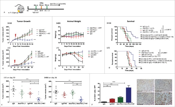
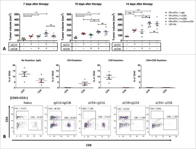
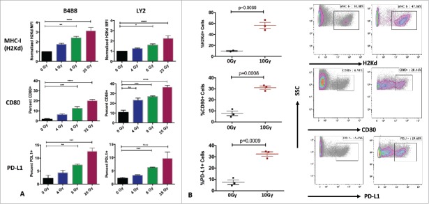
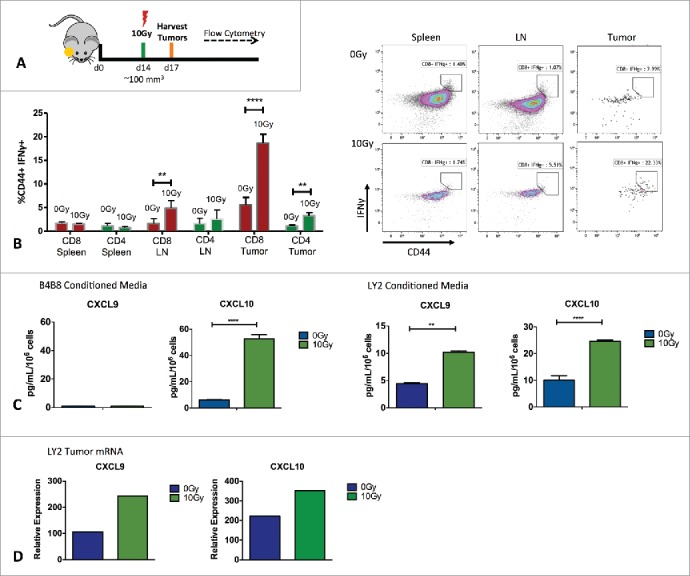
Immunotherapy clinical trials targeting the programmed-death ligand axis (PD-1/PD-L1) show that most head and neck squamous cell carcinoma (HNSCC) patients are resistant to PD-1/PD-L1 inhibition. We investigated whether local radiation to the tumor can transform the immune landscape and render poorly immunogenic HNSCC tumors sensitive to PD-L1 inhibition. We used the first novel orthotopic model of HNSCC with genetically distinct murine cell lines. Tumors were resistant to PD-L1 checkpoint blockade, harbored minimal PD-L1 expression and tumor infiltrating lymphocytes at baseline, and were resistant to radiotherapy. The combination of radiation and PD-L1 inhibition significantly enhanced tumor control and improved survival. This was mediated in part through upregulation of PD-L1 on tumor cells and increased T-cell infiltration after RT, resulting in a highly inflamed tumor. Depletion of both CD4 and CD8 T-cells completely abrogated the effect of anti PD-L1 with radiation on tumor growth. Our findings provide evidence that radiation to the tumor can induce sensitivity to PD-L1 checkpoint blockade in orthotopic models of HNSCC. These findings have direct relevance to high risk HNSCC patients with poorly immunogenic tumors and who may benefit from combined radiation and checkpoint blockade.

Keywords: PD-L1; Radiotherapy; head and neck cancer; immune checkpoint inhibitors.


Figures

Figure 1.
Figure 1.
Analysis of HNSCC tumors in TCGA based on CD8 T cell genomic signature. (A) Patients were selected based on the lower and upper quartile of mRNA expression of CD8, IFNG, PRF1 and GRZMB. Patients in the lower quartile were classified as poorly CD8 infiltrated and patients in the upper quartile were classified as highly CD8 infiltrated. (B) Analysis of the proportion of CD8 T-cells in tumors from each patient group based on CIBERSORT. (C) Analysis of overall survival and disease-free survival in HNSCC patients based on CD8 T-cell profile. Log-ranks hazard ratios are reported.
Figure 2.
Figure 2.
Response of LY2 and B4B8 tumors to treatment with anti PD-L1 monoclonal antibody alone and in combination with RT (single arm RT and IgG were used as control groups). (A) Schematic illustration of treatment schedule. (B) Tumor growth analysis of tumor-bearing mice. Mice received PD-L1 mAb or IgG on day 5 and RT of 10Gy on day 8. PD-L1 or IgG were delivered 2x/week until end of experiment. Statistical analysis was performed on day 20 for LY2 mice and day 36 for B4B8 mice using 2-way analysis of variance (p < 0.0001 in both groups). (C) Average weight of mice in each group (D) Survival analysis of tumor-bearing mice in each group. Hazard ratios were generated based on Log-ranks comparison of the RT+PD-L1 group with each of the other groups. (E) Assessment of tumor growth differences at the last time point when all mice were alive (day 20 for LY2 and day 36 for B4B8). Two-way ANOVA was performed to assess significance. (F) Analysis of tumor-infiltrating lymphocytes 2-weeks after treatment in LY2 mice. Quantification of CD3+ cells was performed by counting the number of positively staining cells per 40x power field. Bars represent SEM from 3–4 independent samples per group. At least 7 fields were quantified per sample. Two-way ANOVA was performed to assess significance.
Figure 3.
Figure 3.
Deciphering the role of T-cell populations in mediating response to RT+anti PD-L1. Mice (n = 5/group) received T-cells depletion antibodies (CD4, CD8 or both) or IgG control 1-week before tumor inoculation. Mice were treated with RT+anti PD-L1 or IgG as described in the methods. Tumor volume was assessed to determine the effect of CD4 and CD8 T-cell depletion on tumor growth.
Figure 4.
Figure 4.
Effects of RT on tumor immunogenicity. (A) Flow cytometric analysis of expression of MHC-I, CD80 and PD-L1 on LY2 and B4B8 HNSCC tumor cells after exposure to increasing doses of RT. Two-way ANOVA was performed to compare statistical significance between the groups .(B) Flow cytometric analysis of the expression of MHC-I, CD80 and PD-L1 on LY2 tumors harvested 72 hours after RT or sham. AquaVi and CD45 staining was performed to gate for live and CD45 negative cells. Bars represent SEM from 3 independent tumor samples. P-values represent significance based on Unpaired T-test analysis.
Figure 5.
Figure 5.
Analysis of T-cell IFNγ activity and in vivo and IFNγ inducible chemokines. (A) Schematic illustration of in vivo experimental setup. (B) Flow cytometric analysis of IFNγ production by activated (CD44+), CD8 and CD4 T-cells from harvested LY2 tumors, spleens and draining lymph nodes. Gating was performed on CD45+, CD3+, live, single cells. Gate assignment was based on FMO and isotype controls. Bars represent SEM from 3 independent experiments with at least 3–4 mice per experimental group. Unpaired T-test was used to assess significance between 0Gy and 10Gy groups. (C) Radiation-induced chemokine production based on ELISA analysis of CXCL9 and CXCL10 in conditioned media from tumor cells irradiated with 10Gy. Conditioned media was harvested 72 hours after irradiation. Unpaired t-test was used for analysis. (C) RT-PCR analysis of CXCL9 and CXCL10 levels in vivo from tumors harvested 72 hours after RT. Experiments were repeated 3 independent times. One representative experiment is shown.

Similar articles

Cited by

References

    1. Parkin DM, Bray F, Ferlay J, Pisani P. Global cancer statistics, 2002. CA Cancer J Clin. 2005;55(2):74-108. doi:10.3322/canjclin.55.2.74. PMID:15761078 - DOI - PubMed
    1. Kamangar F, Dores GM, Anderson WF. Patterns of cancer incidence, mortality, and prevalence across five continents: Defining priorities to reduce cancer disparities in different geographic regions of the world. J Clin Oncol. 2006;24(14):2137-50. doi:10.1200/JCO.2005.05.2308. PMID:16682732 - DOI - PubMed
    1. Taube JM, Klein A, Brahmer JR, Xu H, Pan X, Kim JH, Chen L, Pardoll DM, Topalian SL, Anders RA. Association of PD-1, PD-1 ligands, and other features of the tumor immune microenvironment with response to anti-PD-1 therapy. Clin Cancer Res. 2014;20(19):5064-74. doi:10.1158/1078-0432.CCR-13-3271. PMID:24714771 - DOI - PMC - PubMed
    1. Topalian SL, Sznol M, McDermott DF, Kluger HM, Carvajal RD, Sharfman WH, Brahmer JR, Lawrence DP, Atkins MB, Powderly JD, et al.. Survival, durable tumor remission, and long-term safety in patients with advanced melanoma receiving nivolumab. J Clin Oncol. 2014;32(10):1020-30. doi:10.1200/JCO.2013.53.0105. PMID:24590637 - DOI - PMC - PubMed
    1. Li H, Chiappinelli KB, Guzzetta AA, Easwaran H, Yen RW, Vatapalli R, Topper MJ, Luo J, Connolly RM, Azad NS, et al.. Immune regulation by low doses of the DNA methyltransferase inhibitor 5-azacitidine in common human epithelial cancers. Oncotarget. 2014;5(3):587-98. doi:10.18632/oncotarget.1782. PMID:24583822 - DOI - PMC - PubMed

LinkOut - more resources