

Three dimensional reconstruction of late post traumatic orbital wall defects by customized implants using CAD-CAM, 3D stereolithographic models: A case report
- PMID: 29124002
- PMCID: PMC5670297
- DOI: 10.1016/j.jobcr.2017.09.004
Three dimensional reconstruction of late post traumatic orbital wall defects by customized implants using CAD-CAM, 3D stereolithographic models: A case report
Abstract
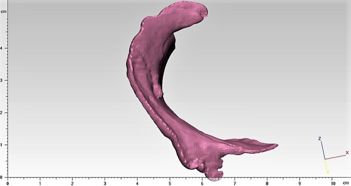
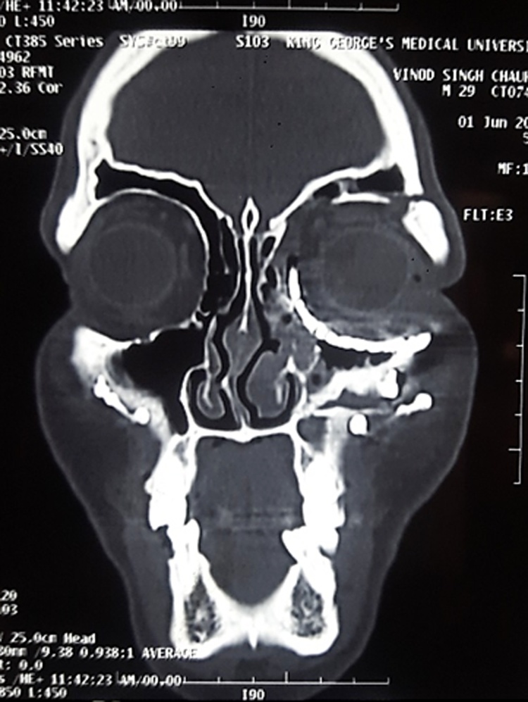
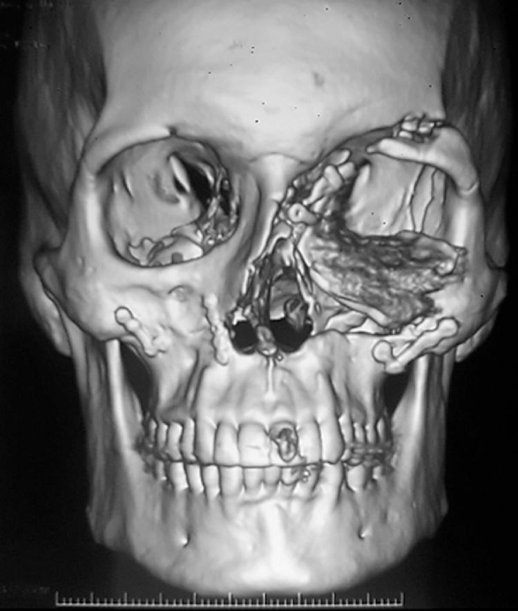
Aim: Purpose of this case report is to highlight the precision and accuracy obtained with patient specific implants for orbital reconstruction designed on the basis of volumetric analysis of orbital computed tomographic scan (CT) scans using virtual planning, computerised designing and manufacturing and stereolithographic models to correct late post-traumatic orbital deformities such as enophthalmos and diplopia.
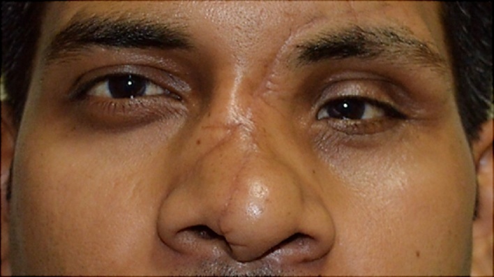
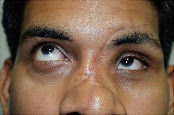
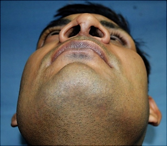
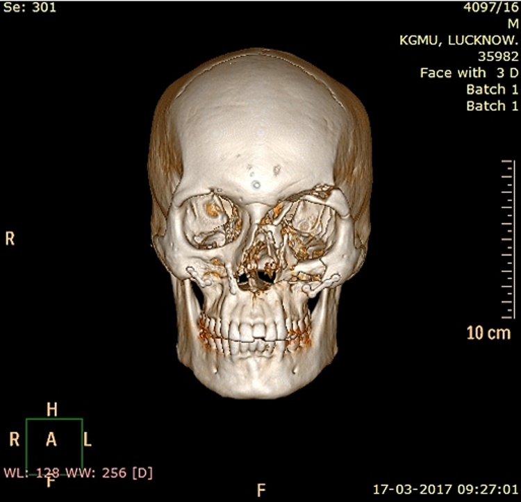
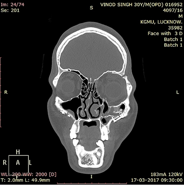
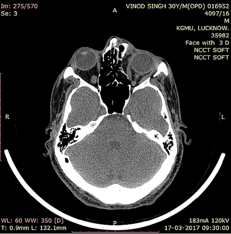
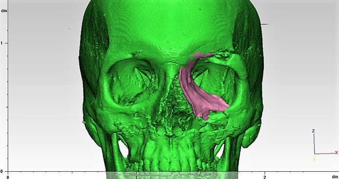
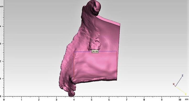
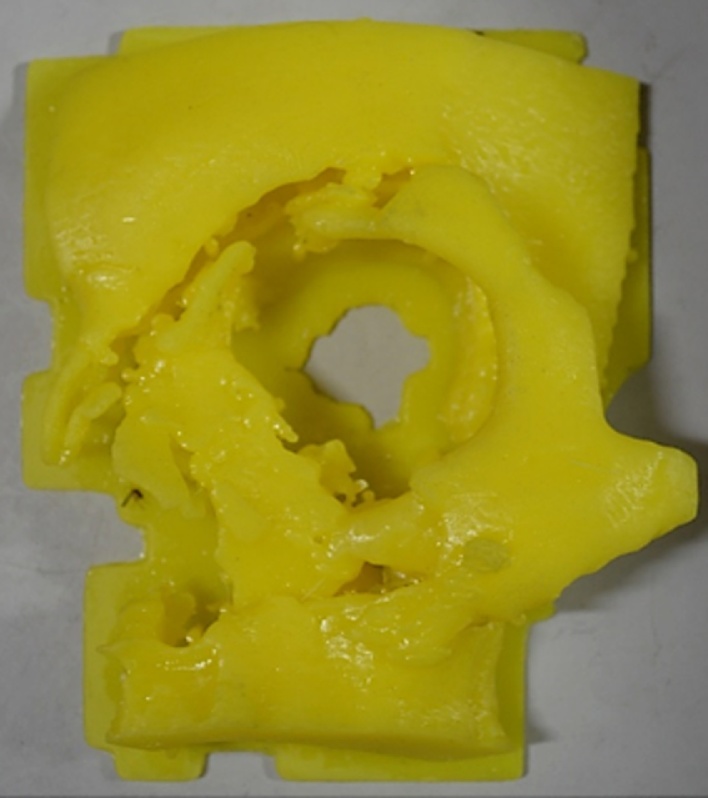
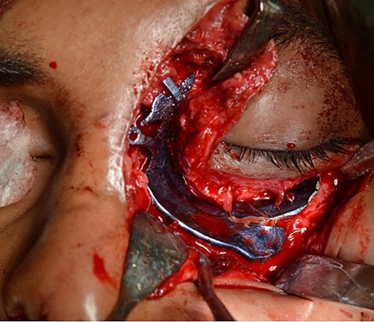
Material and methods: This case report describes a patient who visited our outpatient clinic for correction of enophthalmos and persistent diplopia in upward gaze, seven months post trauma. Three dimensional (3D) virtual treatment planning was carried out by using the 3D CT data. The unaffected orbit of the contralateral side was superimposed on the deformed orbit to highlight the defect and a customized implant was designed in the desired size and shape on the virtual model using computer aided designing and manufacturing (CAD-CAM) and milled in titanium mesh for precise anatomic orbital reconstruction.
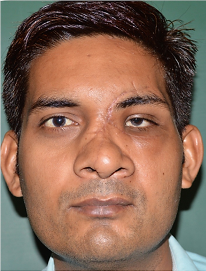
Results: There was a marked improvement in both the diplopia in upward gaze and enophthalmos post surgery when the customized patient specific orbital implant was used.
Conclusion: The concept of using customized implant with the help of 3D virtual treatment planning, 3D stereolithographic models and CAD-CAM greatly improves the correction of extremely difficult late post-traumatic orbital deformities.
Keywords: Customized implant; Diplopia; Enophthalmos; Orbital volume.
Figures

References
-
- Pedemonte C., Sáez F., Vargas I., González L.E., Canales M., Salazar K. Can customized implants correct enophthalmos and delayed diplopia in posttraumatic orbital deformities? A volumetric analysis. Int J Oral Maxillofac Surg. 2016;45:1086–1094. - PubMed
-
- Wolff J., Sándor G.K., Pyysalo M., Miettinen A., Koivumäki A.V., Kainulainen V.T. Late reconstruction of orbital and naso-orbital deformities. Oral Maxillofacial Surg Clin N Am. 2013;25:683–695. - PubMed
-
- Baumann A., Sinko K., Dorner G. Late reconstruction of the orbit with patient-specific implants using computer-aided planning and navigation. J Oral Maxillofac Surg. 2015;73:S101–S106. - PubMed
-
- An J.G., Zhang Y., Zhang Z.Y. Computer-assisted fabricated individual titanium mesh for reconstruction of orbital wall. Beijing Da Xue Xue Bao. 2008;40(1):88–91. - PubMed
-
- Metzger M.C., Schön R., Weyer N. Anatomical 3-dimensional pre-bent titanium implant for orbital floor fractures. Ophthalmology. 2006;113(10):1863–1868. - PubMed
LinkOut - more resources
Full Text Sources
Other Literature Sources
Miscellaneous
