. 2015 Mar 24:1:33-51.
doi: 10.1016/j.bbrep.2015.03.003. eCollection 2015 May.

Intrinsically disordered proteins in the nucleus of human cells

Affiliations

Intrinsically disordered proteins in the nucleus of human cells

Telma Frege et al. Biochem Biophys Rep. .

Abstract

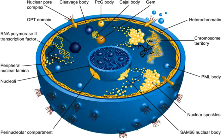
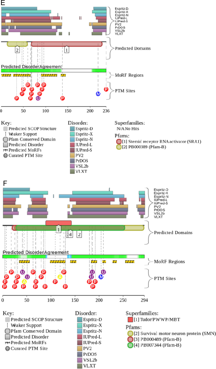
Intrinsically disordered proteins are known to perform a variety of important functions such as macromolecular recognition, promiscuous binding, and signaling. They are crucial players in various cellular pathway and processes, where they often have key regulatory roles. Among vital cellular processes intimately linked to the intrinsically disordered proteins is transcription, an intricate biological performance predominantly developing inside the cell nucleus. With this work, we gathered information about proteins that exist in various compartments and sub-nuclear bodies of the nucleus of the human cells, with the goal of identifying which ones are highly disordered and which functions are ascribed to the disordered nuclear proteins.

Keywords: Cell nucleus; Homo sapiens; Intrinsically disordered proteins; Nuclear compartments; Nuclear proteins; Protein function.


Figures

Fig. 1
Fig. 1
Sub-nuclear compartments. Reproduced with the permission from .
Fig. 2
Fig. 2
Peculiarities of disorder distribution in human nuclear proteins.
Fig. 3
Fig. 3
Detailed representation of the disorder distribution in various nuclear compartments. This bar graph shows how many proteins in each compartment have disordered regions accounting for ≥0%, ≥10%, ≥20%, …, ≥80%, ≥90%, and 100% of their residues.
Fig. 4
Fig. 4
Evaluating intrinsic disorder in human nuclear proteins by combined binary disorder classifiers, CH-plot and CDF , , . Here, the coordinates of each point were calculated as a distance of the corresponding protein in the CH-plot from the boundary (Y-coordinate) and an average distance of the respective CDF curve from the CDF boundary (X-coordinate). The four quadrants correspond to the following predictions: Q1, proteins predicted to be disordered by CH-plots, but ordered by CDFs; Q2, ordered proteins; Q3, proteins predicted to be disordered by CDFs, but compact by CH-plots (i.e., putative molten globules or hybrid proteins); Q4, proteins predicted to be disordered by both methods.
Fig. 5
Fig. 5
Abundance of intrinsic disorder in human nuclear proteins. JRONN vs. PONDR® VSL2B plot representing the correlation between the disorder content evaluated by PONDR® VSL2B (x-axis) , and by JRONN (y-axis) . Solid black line corresponds to the diagonal. Dashed line shows linear fit of all the data point to the equation (R2=0.90):.DSJRONN=1.4±1+0.85±0.02×DSPONDR. Following the accepted practice, two arbitrary cutoffs for the levels of intrinsic disorder are used to classify proteins as highly ordered (IDP score<10% light blue field), moderately disordered (10%≤IDP score<30%, light yellow field) and highly disordered (IDP score≥30%, light pink field) .
Fig. 6
Fig. 6
Analysis of the interactivity of the several human nuclear proteins by the STRING platform . Analyzed proteins are: (A) KI-67 (UniProt ID: P46013), (B) N-CoR2 (UniProt ID: Q9Y618), (C) BRCA1 (UniProt ID: P38398), (D). SON (UniProt ID: P18583), (E) ATRX (UniProt ID: P46100), and (F) RLP24 (UniProt ID: Q9UHA3). STRING produces the network of predicted associations for a particular group of proteins. The network nodes are proteins, whereas the edges represent the predicted or known functional associations. An edge is drawn with up to 7 differently colored lines that represent the existence of the seven types of evidence used in predicting the associations. A red line indicates the presence of fusion evidence; a green line – neighborhood evidence; a blue line – co-occurrence evidence; a purple line – experimental evidence; a yellow line – text mining evidence; a light blue line – database evidence; a black line – co-expression evidence .
Fig. 7
Fig. 7
Evaluation of the functional intrinsic disorder propensity of representative human nuclear proteins by D2P2 database (http://d2p2.pro/) . (A) Sp1 (UniProt ID: P08047). (B) Sam68 (UniProt ID: Q07666). (C) CASC3 (UniProt ID: O15234). (D) Coilin (UniProt ID: P38432). (E) SRA1p (UniProt ID: Q9HD15). (F) SMN (UniProt ID: Q16637).
Fig. 7
Fig. 7
Evaluation of the functional intrinsic disorder propensity of representative human nuclear proteins by D2P2 database (http://d2p2.pro/) . (A) Sp1 (UniProt ID: P08047). (B) Sam68 (UniProt ID: Q07666). (C) CASC3 (UniProt ID: O15234). (D) Coilin (UniProt ID: P38432). (E) SRA1p (UniProt ID: Q9HD15). (F) SMN (UniProt ID: Q16637).

References

    1. Goodsell D.S. Miniseries: illustrating the machinery of life: eukaryotic cell panorama. Biochem. Mol. Biol. Educ.: Bimon. Publ. Int. Union Biochem. Mol. Biol. 2011;39:91–101. - PubMed
    1. Tripathi V., Prasanth K.V. John Wiley & Sons Ltd.; Chichester: 2011. Cell Nucleus. eLS.
    1. Hancock R., Jeon K., editors. Academic Press; Amsterdam, Boston, Heidelberg, London, New York, Oxford, Paris, San Diego, San Francisco, Singapoe, Sydney, Tokyo: 2014. New Models of the Cell Nucleus: Crowding, Entropic Forces, Phase Separation, and Fractals.
    1. Jevtic P., Edens L.J., Vukovic L.D., Levy D.L. Sizing and shaping the nucleus: mechanisms and significance. Curr. Opin. Cell Biol. 2014;28:16–27. - PMC - PubMed
    1. Fakan S. The functional architecture of the nucleus as analysed by ultrastructural cytochemistry. Histochem. Cell Biol. 2004;122:83–93. - PubMed