

A method of gentle hydration to prepare oil-free giant unilamellar vesicles that can confine enzymatic reactions
- PMID: 29124169
- PMCID: PMC5668676
- DOI: 10.1016/j.bbrep.2015.07.005
A method of gentle hydration to prepare oil-free giant unilamellar vesicles that can confine enzymatic reactions
Abstract
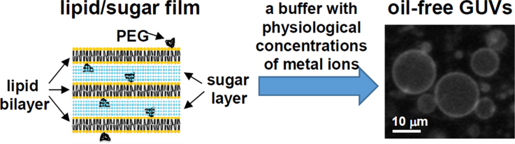
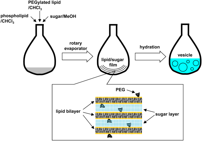
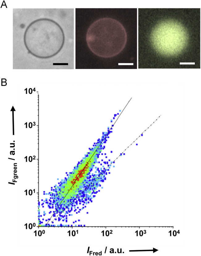
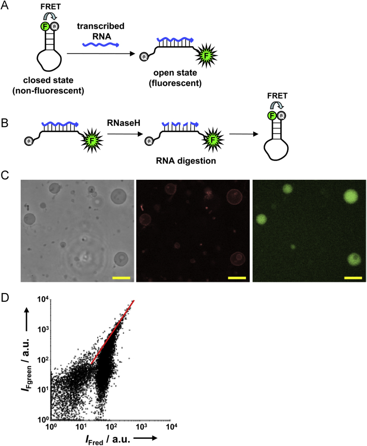
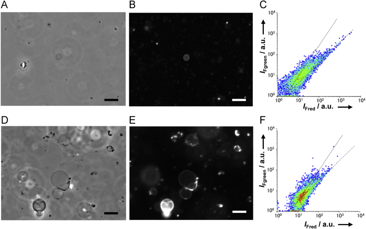
We report a new and improved method to prepare, by gentle hydration of lipid films, oil-free giant unilamellar vesicles (GUVs), in which enzymatic reactions can be encapsulated. The traditional method of gentle hydration requires very low concentrations of metal ions, whereas enzymatic reactions generally require mono- and divalent metal ions at physiological concentrations. In order to improve the production of oil-free GUVs that can confine enzymatic reactions, we developed a novel method also based on gentle hydration, but in which the precursor lipid film was doped with both 1,2-dioleoyl-sn-glycero-3-phosphoethanolamine-N-[methoxy(polyethylene glycol)-2000] (PEGylated lipid) and sugar. Close examination of the size, shape, and lamellarity of vesicles prepared in this manner demonstrated that the process improves the production of oil-free GUVs even at low temperatures and physiological salt concentrations. PEGylated lipid and sugar were found to synergistically improve GUV formation. Finally, we demonstrate the successful enzymatic synthesis of RNA within oil-free GUVs that were prepared on ice.
Keywords: Flow cytometry; GMV, giant multilamellar vesicle; GUV; GUV, giant unilamellar vesicle; Gentle hydration; Liposome; Membrane protein; Nile red, 9-(diethylamino)-5 H-benzo(α)phenoxazin-5-one; PEGylated lipid, 1,2-dioleoyl-sn-glycero-3-phosphoethanolamine-N-[methoxy(polyethylene glycol)-2000]; PSGH, PEGylated-lipid-and-sugar-doped gentle hydration; Synthetic biology.
Figures
Similar articles
-
Efficient formation of giant liposomes through the gentle hydration of phosphatidylcholine films doped with sugar.Colloids Surf B Biointerfaces. 2009 Jan 1;68(1):98-105. doi: 10.1016/j.colsurfb.2008.09.023. Epub 2008 Oct 2. Colloids Surf B Biointerfaces. 2009. PMID: 18993037
-
Dynamics of giant vesicle assembly from thin lipid films.J Colloid Interface Sci. 2024 May;661:1033-1045. doi: 10.1016/j.jcis.2024.02.022. Epub 2024 Feb 5. J Colloid Interface Sci. 2024. PMID: 38335788
-
Evaluation of dextran(ethylene glycol) hydrogel films for giant unilamellar lipid vesicle production and their application for the encapsulation of polymersomes.Soft Matter. 2017 Aug 23;13(33):5580-5588. doi: 10.1039/c7sm00551b. Soft Matter. 2017. PMID: 28730206 Free PMC article.
-
Lipid-polymer hybrid nanoparticles as a new generation therapeutic delivery platform: a review.Eur J Pharm Biopharm. 2013 Nov;85(3 Pt A):427-43. doi: 10.1016/j.ejpb.2013.07.002. Epub 2013 Jul 17. Eur J Pharm Biopharm. 2013. PMID: 23872180 Review.
-
Giant unilamellar vesicles - a perfect tool to visualize phase separation and lipid rafts in model systems.Acta Biochim Pol. 2009;56(1):33-9. Epub 2009 Mar 17. Acta Biochim Pol. 2009. PMID: 19287805 Review.
Cited by
-
Rapid Multi-Well Evaluation of Assorted Materials for Hydrogel-Assisted Giant Unilamellar Vesicle Production: Empowering Bottom-Up Synthetic Biology.Gels. 2025 Jan 2;11(1):29. doi: 10.3390/gels11010029. Gels. 2025. PMID: 39852000 Free PMC article.
-
Controlled Dendrimersome Nanoreactor System for Localized Hypochlorite-Induced Killing of Bacteria.ACS Nano. 2020 Dec 22;14(12):17333-17353. doi: 10.1021/acsnano.0c07459. Epub 2020 Dec 8. ACS Nano. 2020. PMID: 33290039 Free PMC article.
-
Tuning of TRAIL clustering on the surface of nanoscale liposomes by phase separation.Nanoscale Adv. 2023 Dec 29;6(2):402-405. doi: 10.1039/d3na00841j. eCollection 2024 Jan 16. Nanoscale Adv. 2023. PMID: 38235079 Free PMC article.
References
-
- Walde P., Cosentino K., Engel H., Stano P. Giant vesicles: preparations and applications. ChemBioChem. 2010;11:848–865. - PubMed
-
- Kahya N. Protein–protein and protein–lipid interactions in domain-assembly: lessons from giant unilamellar vesicles. Biochim. Biophys. Acta. 2010;1798:1392–1398. - PubMed
-
- Szostak J.W., Bartel D.P., Luisi P.L. Synthesizing life. Nature. 2001;409:387–390. - PubMed
-
- Maeda Y.T., Nakadai T., Shin J., Uryu K., Noireaux V., Libchaber A. Assembly of MreB filaments on liposome membrane: a synthetic biology approach. ACS Synth. Biol. 2012;1:53–59. - PubMed
LinkOut - more resources
Full Text Sources
Other Literature Sources
