. 2018 Jan;33(1):136-145.
doi: 10.1002/mds.27217. Epub 2017 Nov 10.

Dysfunction in emotion processing underlies functional (psychogenic) dystonia

Affiliations

Dysfunction in emotion processing underlies functional (psychogenic) dystonia

Alberto J Espay et al. Mov Disord. 2018 Jan.

Abstract

Objective: We sought to determine whether abnormalities in emotion processing underlie functional (psychogenic) dystonia, one of the most common functional movement disorders.

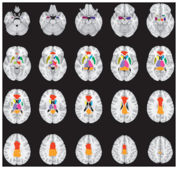
Methods: Motor and emotion circuits were examined in 12 participants with functional dystonia, 12 with primary organic dystonia, and 25 healthy controls using functional magnetic resonance imaging at 4T and a finger-tapping task (motor task), a basic emotion-recognition task (emotional faces task), and an intense-emotion stimuli task.

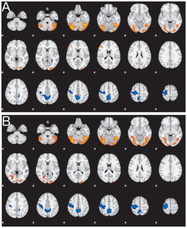
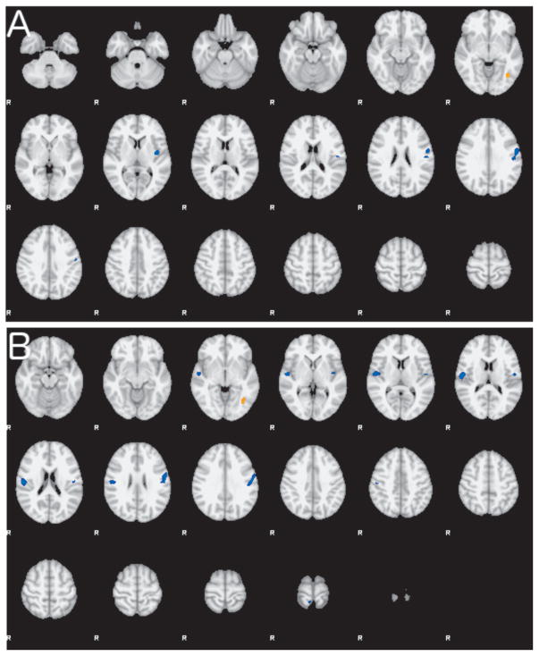
Results: There were no differences in motor task activation between groups. In the faces task, when compared with the other groups, functional dystonia patients showed areas of decreased activation in the right middle temporal gyrus and bilateral precuneus and increased activation in the right inferior frontal gyrus, bilateral occipital cortex and fusiform gyrus, and bilateral cerebellum. In the intense-emotion task, when compared with the other groups, functional dystonia patients showed decreased activation in the left insular and left motor cortices (compared to organic dystonia, they showed an additional decrease in activation in the right opercular cortex and right motor cortex) and increased activation in the left fusiform gyrus.

Conclusions: Functional dystonia patients exhibited stimulus-dependent altered activation in networks involved in motor preparation and execution, spatial cognition, and attentional control. These results support the presence of network dysfunction in functional dystonia. © 2017 International Parkinson and Movement Disorder Society.

Keywords: Functional dystonia; conversion disorder; emotion processing; fMRI; functional movement disorders; psychogenic dystonia.


Conflict of interest statement

Conflicts of Interest/Financial Disclosures over last 24 months:

Dr. Espay has received grant support from NIH (NIMH, 1K23MH092735), Great Lakes Neurotechnologies and the Michael J Fox Foundation; personal compensation as a consultant/scientific advisory board member for Abbvie, TEVA, Impax, Merz, Acadia, Cynapsus, Lundbeck, and USWorldMeds; publishing royalties from Lippincott Williams & Wilkins, Cambridge University Press, and Springer; and honoraria from Abbvie, UCB, USWorldMeds, Lundbeck, Acadia, the American Academy of Neurology, and the Movement Disorders Society.

Dr. Vannest receives funding from the NIH.

Mr. Maloney Norris, Dr. Eliassen, and Ms. Neefus have nothing to disclose.

Dr. Allendorfer has received funding from the Shor Foundation for Epilepsy Research. She serves as an associate editor of the journal Restorative Neurology and Neuroscience.

Dr. Chen received honoraria from Allergan, and Merz, Ipsen and UCB. He is the Editor-in- Chief of the Canadian Journal of Neurological Sciences. He received research grants from the Canadian Institutes of Health Research, Michael J. Fox Foundation for Parkinson Research, Medtronic Inc, the Weston Foundation and Merz Pharma.

Dr. Szaflarski has received research funding from NIH, Shor Foundation for Epilepsy Research, Eisai, Epilepsy Foundation of America, Food and Drug Administration, Compumedics Neuroscan, Inc., Department of Defense, State of Alabama, and the University of Alabama at Birmingham (UAB). While this research was conducted, he was supported by NIH K23 NS052468. He serves as an associate editor of the journals Restorative Neurology and Neuroscience and Journal of Epileptology and on editorial boards of the journals Epilepsy & Behavior, Folia Medica Copernicana and Journal of Medical Science.

Figures

Figure 1
Figure 1. Atlas-based subcortical regions of interest selected for connectivity analysis
The anatomical regions of interest included the anterior cingulate gyrus, posterior cingulate gyrus and left and right regions of the uncus/amygdala, thalamus, caudate, and putamen from the Harvard-Oxford Subcortical Atlas and left and right subthalamic nuclei from the Subthalamic Nucleus atlas.
Figure 2
Figure 2. Group differences in the Emotional Faces task-related activation
A. Composite map shows the differences in activation between FD and HC during the Faces task contrasting faces versus a fixation cross as control. Regions of greater activity in FD are shown in red-yellow hues; regions of lower activity in FD in blue hues. B. Composite map showing the differences in activation between FD and OD during the Faces task contrasting faces versus a fixation cross as control. Regions of greater activity in FD are shown in red-yellow hues; regions of lower activity in FD in blue hues. Both group contrasts are thresholded at Z > 2.3 and corrected p<0.05.
Figure 3
Figure 3. Group differences in the CPT-END task-related activation
A. Composite map shows the differences in activation between FD and HC during the CPT-END task contrasting intense emotional images versus squares as control. Regions of lower activity in FD are shown in blue hues and a small region of greater activity in FD is shown in orange. B. Composite map showing differences in activation between FD and OD during the CPT-END task contrasting intense emotional images versus squares. Regions of lower activity in FD are shown in blue hues. Both group contrasts are thresholded at Z > 2.3 and corrected p<0.05.

References

    1. Fahn S, Williams DT. Psychogenic dystonia. Adv Neurol. 1988;50:431–455. - PubMed
    1. Espay AJ, Lang AE. Phenotype-specific diagnosis of functional (psychogenic) movement disorders. Curr Neurol Neurosci Rep. 2015;15:556. - PubMed
    1. Nowak DA, Fink GR. Psychogenic movement disorders: aetiology, phenomenology, neuroanatomical correlates and therapeutic approaches. Neuroimage. 2009;47:1015–1025. - PubMed
    1. Schrag AE, Mehta AR, Bhatia KP, et al. The functional neuroimaging correlates of psychogenic versus organic dystonia. Brain. 2013;136:770–781. - PMC - PubMed
    1. Voon V, Brezing C, Gallea C, et al. Emotional stimuli and motor conversion disorder. Brain. 2010;133:1526–1536. - PMC - PubMed

Publication types

MeSH terms