Review
. 2018 Jan;12(1):3-20.
doi: 10.1002/1878-0261.12155. Epub 2017 Nov 27.

Emerging functions of the EGFR in cancer

Affiliations
Review

Emerging functions of the EGFR in cancer

Sara Sigismund et al. Mol Oncol. 2018 Jan.

Abstract

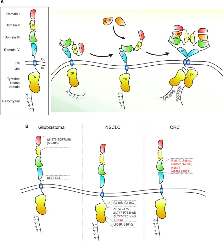
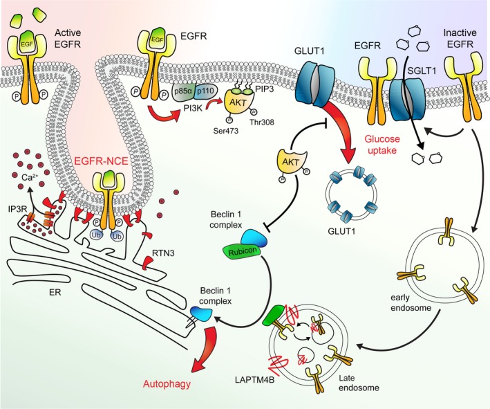
The physiological function of the epidermal growth factor receptor (EGFR) is to regulate epithelial tissue development and homeostasis. In pathological settings, mostly in lung and breast cancer and in glioblastoma, the EGFR is a driver of tumorigenesis. Inappropriate activation of the EGFR in cancer mainly results from amplification and point mutations at the genomic locus, but transcriptional upregulation or ligand overproduction due to autocrine/paracrine mechanisms has also been described. Moreover, the EGFR is increasingly recognized as a biomarker of resistance in tumors, as its amplification or secondary mutations have been found to arise under drug pressure. This evidence, in addition to the prominent function that this receptor plays in normal epithelia, has prompted intense investigations into the role of the EGFR both at physiological and at pathological level. Despite the large body of knowledge obtained over the last two decades, previously unrecognized (herein defined as 'noncanonical') functions of the EGFR are currently emerging. Here, we will initially review the canonical ligand-induced EGFR signaling pathway, with particular emphasis to its regulation by endocytosis and subversion in human tumors. We will then focus on the most recent advances in uncovering noncanonical EGFR functions in stress-induced trafficking, autophagy, and energy metabolism, with a perspective on future therapeutic applications.

Keywords: EGFR; cancer; membrane trafficking; signal transduction.


Figures

Figure 1
Figure 1
Scheme of EGFR and its mutations in glioblastoma and in lung and colorectal cancer. (A) Schematic representation of the EGFR and EGF‐induced receptor activation. The EGFR extracellular region encompasses domains I, II, III, and IV; following are the transmembrane region (TM), the intracellular juxtamembrane domain (iJM), the tyrosine kinase domain (TK), and the carboxyl‐terminal tail (carboxy tail). EGF binding to the receptor unmasks a dimerization motif and determines structural rearrangements that are conveyed to the cytoplasmic domain allowing the formation of asymmetric dimers between the two juxtaposed catalytic domains. (B) Most frequent EGFR mutations in glioblastoma, in NSCLC (non‐small‐cell lung cancer), and in CRC (colorectal cancer). Mutations found in tumors resistant to EGFR blockade are shown in red. In CRC, the indicated EGFR mutations have been identified in patients that progressed upon cetuximab treatment (Arena et al., 2015, 2016; Montagut et al., 2012; Van Emburgh et al., 2016).
Figure 2
Figure 2
Active and inactive EGFR‐related functions. This picture schematizes some noncanonical EGFR functions. From left to right: EGFR stimulated with high EGF doses (active EGFR) is phosphorylated (P) and ubiquitinated (Ub) and undergoes both clathrin‐mediated endocytosis (not depicted) and nonclathrin‐dependent endocytosis (NCE), the latter dependent on the formation of RTN3‐mediated ERPM contact sites. This is accompanied by calcium release in the proximity of contact sites, which likely controls fission of the tubular invagination. It is still unclear whether RTN3 is the tethering factor between the ER and the PM (as depicted), or it is just involved the tubulation of cortical ER, but not directly engaged at contact sites. EGFR ligand stimulation elicits the classical signaling cascade based on the recruitment of PI3K (made of its p85 regulatory subunit and p110 catalytic subunit) that catalyzes the formation of PIP3s. PIP3s bind to the PH domain of AKT and of phosphoinositide‐dependent kinase‐1, PDK1. PDK1 phosphorylates AKT on Thr308, while mammalian target of rapamycin complex 2, mTORC2 (not depicted here), is responsible for phosphorylation on Ser 473, leading to full AKT activation. Active AKT inhibits autophagy and blocks GLUT1 endocytosis. This latter function leads to higher levels of GLUT1 at the plasma membrane, increasing the uptake of glucose. In addition, ligand‐independent direct interaction of EGFR (inactive EGFR) and SGLT1 stabilizes the glucose transporter at the cell surface promoting high glucose uptake. Ligand‐unbound EGFR constitutively internalizes into early and late endosomes where it is sequestered by LAPTM4B. Here, the inactive EGFR interacts with Rubicon causing its dissociation from Beclin‐1. Beclin‐1 complex can now initiates autophagy on the ER membrane.

References

    1. Aguet F, Antonescu CN, Mettlen M, Schmid SL and Danuser G (2013) Advances in analysis of low signal‐to‐noise images link dynamin and AP2 to the functions of an endocytic checkpoint. Dev Cell 26, 279–291. - PMC - PubMed
    1. Arena S, Bellosillo B, Siravegna G, Martínez A, Cañadas I, Lazzari L, Ferruz N, Russo M, Misale S, González I et al (2015) Emergence of multiple EGFR extracellular mutations during cetuximab treatment in colorectal cancer. Clin Cancer Res 21, 2157–2166. - PubMed
    1. Arena S, Siravegna G, Mussolin B, Kearns JD, Wolf BB, Misale S, Lazzari L, Bertotti A, Trusolino L, Adjei AA et al (2016) MM‐151 overcomes acquired resistance to cetuximab and panitumumab in colorectal cancers harboring EGFR extracellular domain mutations. Sci Transl Med 8, 324ra314. - PubMed
    1. Arteaga CL, Ramsey TT, Shawver LK and Guyer CA (1997) Unliganded epidermal growth factor receptor dimerization induced by direct interaction of quinazolines with the ATP binding site. J Biol Chem 272, 23247–23254. - PubMed
    1. Babic I, Anderson ES, Tanaka K, Guo D, Masui K, Li B, Zhu S, Gu Y, Villa GR, Akhavan D et al (2013) EGFR mutation‐induced alternative splicing of Max contributes to growth of glycolytic tumors in brain cancer. Cell Metab 17, 1000–1008. - PMC - PubMed

Publication types