Review
. 2017;24(sup1):94-107.
doi: 10.1080/10717544.2017.1399300.

Mesoporous carbon nanomaterials in drug delivery and biomedical application

Affiliations
Review

Mesoporous carbon nanomaterials in drug delivery and biomedical application

Qinfu Zhao et al. Drug Deliv. 2017.

Abstract

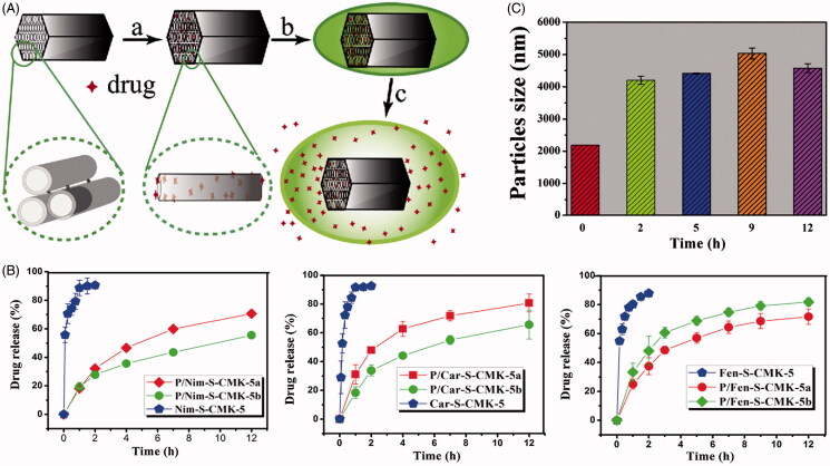
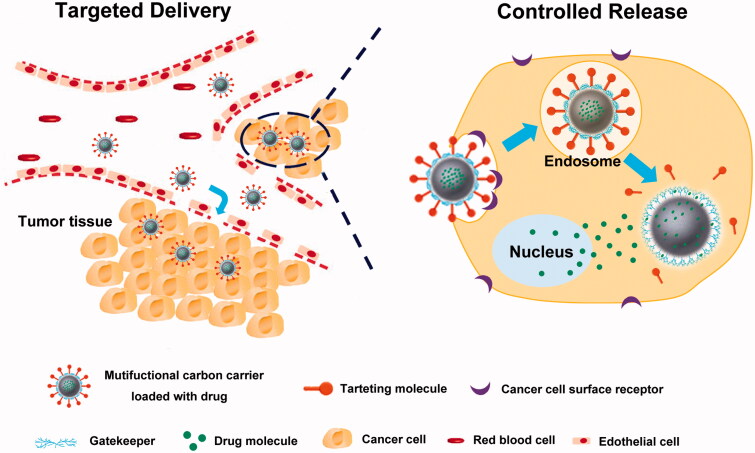
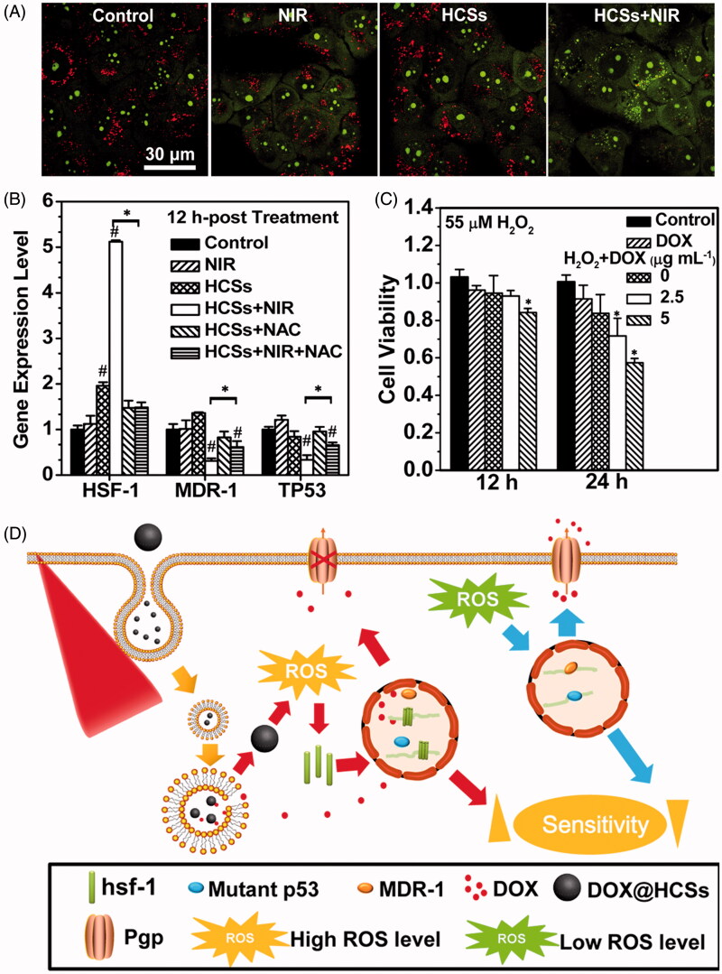
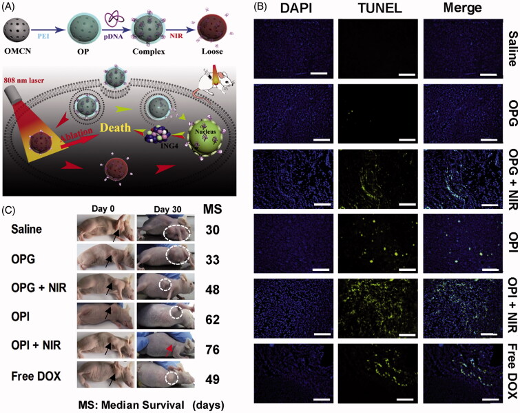
Recent development of nano-technology provides highly efficient and versatile treatment methods to achieve better therapeutic efficacy and lower side effects of malignant cancer. The exploration of drug delivery systems (DDSs) based on nano-material shows great promise in translating nano-technology to clinical use to benefit patients. As an emerging inorganic nanomaterial, mesoporous carbon nanomaterials (MCNs) possess both the mesoporous structure and the carbonaceous composition, endowing them with superior nature compared with mesoporous silica nanomaterials and other carbon-based materials, such as carbon nanotube, graphene and fullerene. In this review, we highlighted the cutting-edge progress of carbon nanomaterials as drug delivery systems (DDSs), including immediate/sustained drug delivery systems and controlled/targeted drug delivery systems. In addition, several representative biomedical applications of mesoporous carbon such as (1) photo-chemo synergistic therapy; (2) delivery of therapeutic biomolecule and (3) in vivo bioimaging are discussed and integrated. Finally, potential challenges and outlook for future development of mesoporous carbon in biomedical fields have been discussed in detail.

Keywords: Mesoporous carbon nanomaterials; biocompatibility; biomedical applications; drug delivery systems; photothermal therapy.


Conflict of interest statement

The authors report no conflicts of interest.

Figures

Figure 1.
Figure 1.
(A) Schematic diagram of the drug loading and release process of PDDA coated S-CMK-5 composites. (a) Drug solution is filled into the interior of S-CMK-5 through ultrasonication; (b) drug-loaded S-CMK-5 is added to the PDDA solution and the synthesis of composites is carried out through electrostatic interaction; (c) drug is released from the swelling PDDA coated S-CMK-5 in the release medium. (B) Nimodipine, carvedilol and fenofibrate release profiles of different drug release systems. (C) Particles size changes for P/S-CMK-5 at different sampling times in enzyme-free simulated gastric fluid (SGF, pH 1.2) (Reproduced with permission from Zhang et al., , copyright by Elsevier).
Figure 2.
Figure 2.
Schematic illustration for the in vivo process of controlled and targeted drug delivery system based on MCNs.
Figure 3.
Figure 3.
(A) Changes in the lysosomal membrane permeation induced by the photothermal effects determined by AO staining. (B) Impact of intracellular ROS levels on the expression of HSF-1, MDR-1 and TP53 genes. The asterisk (*) shows significant differences between test samples and the sample under laser irradiation (HCSs + NIR) (p < .05); the pound symbol (#) indicates significant differences between test samples and the control. (C) Change in the DOX sensitivity of MCF-7/ADR cells treated with 55 μM H2O2. Asterisk (*) indicates significant differences between control and test samples. (D) Combatting the chemotherapeutic resistance of cancer using HCSs under NIR laser irradiation. (Reprinted with permission from (Wang et al., 2015c), copyright 2015 American Chemical Society).
Figure 4.
Figure 4.
(A) Illustration of the synthesis and combined photothermal combined gene therapy achieved by PEI-grafted OMCN. (B) Apoptosis results on 14th day postinjection with different treatments based on TUNEL assay. Blue: DAPI-stained nucleus; green: FITC-labeled apoptosis cells. (C) Images of mice on 30th day postinjection and median survival data with different treatments. Black arrows and white dotted circles indicated the tumor sites before and after treatments, respectively. (Reproduced with permission from (Meng et al., 2016), copyright by Elsevier).
Figure 5.
Figure 5.
(A) Schematic illustration of the sensing principle based on the OMCN/P0-Cy3 aptasensor. (B) MUC1 responsive fluorescent recovery in the buffer solution recorded by fluorescence spectrophotometer. (C–E) Sensing performances of the OMCN/P0-Cy3 aptasensor in cell level (confocal fluorescence microscopy image), tissue level (inverted fluorescence microscopy image), and whole animal level (in vivo fluorescent image), respectively. (Reprinted with permission from (Li et al., 2015a), copyright 2015 American Chemical Society).

Similar articles

Cited by

References

    1. Ai K, Liu Y, Ruan C, et al. (2013). Sp2 C-dominant N-doped carbon sub-micrometer spheres with a tunable size: a versatile platform for highly efficient oxygen-reduction catalysts. Adv Mater 25:998–1003. - PubMed
    1. Akhavan O, Ghaderi E. (2013). Graphene nanomesh promises extremely efficient in vivo photothermal therapy. Small 9:3593–601. - PubMed
    1. Alhmoud H, Delalat B, Elnathan R, et al. (2015). Porous silicon nanodiscs for targeted drug delivery. Adv Funct Mater 25:1137–45.
    1. Bai L, Zhao Q, Wang J, et al. (2015). Mechanism study on pH-responsive cyclodextrin capped mesoporous silica: effect of different stalk densities and the type of cyclodextrin. Nanotechnology 26:165704. - PubMed
    1. Boesch D, Gavériaux C, Jachez B, et al. (1991). In vivo circumvention of P-glycoprotein-mediated multidrug resistance of tumor cells with SDZ PSC 833. Cancer Res 51:4226–33. - PubMed