

Wingspan stent delivery catheter fracture and the TRAP technique for endovascular salvage
- PMID: 29125024
- PMCID: PMC5772540
- DOI: 10.1177/1591019917737734
Wingspan stent delivery catheter fracture and the TRAP technique for endovascular salvage
Abstract
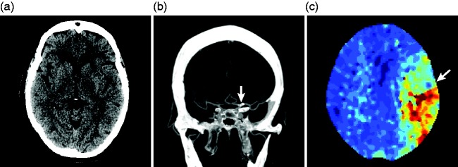
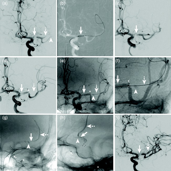
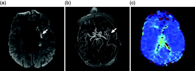
Background Intracranial atherosclerotic disease may result in ischemic infarction and has a high rate of recurrent ischemic strokes despite medical therapy. Patients who fail medical therapy may undergo endovascular treatment with cerebral artery angioplasty and possible Wingspan stent placement. We present a unique case of Wingspan delivery microcatheter fracture that resulted in a retained foreign body and an endovascular salvage maneuver. Case description An elderly patient presented with an acute ischemic stroke due to a severe stenosis in the proximal left middle cerebral artery (MCA). The patient failed non-invasive medical treatment and underwent endovascular treatment with angioplasty and Wingspan stent placement. Following Wingspan stent deployment, the stent delivery catheter fractured, and the retained catheter fragment resulted in MCA occlusion. The foreign body was retrieved by balloon catheter inflation within an intermediate catheter adjacent to the proximal end of the fractured catheter and removal of the entire construct (TRAP technique). Conclusions Wingspan delivery microcatheter fracture is a rare event. The TRAP technique may be used for successful retrieval of a retained foreign body.
Keywords: Stroke; Wingspan; angioplasty; foreign body; intracranial atherosclerotic disease.
Figures
Similar articles
-
Long-term Outcome of Angioplasty Using a Wingspan Stent, Post-Stent Balloon Dilation and Aggressive Restenosis Management for Intracranial Arterial Stenosis.Clin Neuroradiol. 2020 Mar;30(1):159-169. doi: 10.1007/s00062-019-00793-1. Epub 2019 May 23. Clin Neuroradiol. 2020. PMID: 31123775
-
Safety and Efficacy of Wingspan Stenting for Severe Symptomatic Atherosclerotic Stenosis of the Middle Cerebral Artery: Analysis of 278 Continuous Cases.J Stroke Cerebrovasc Dis. 2016 Oct;25(10):2368-72. doi: 10.1016/j.jstrokecerebrovasdis.2016.05.035. Epub 2016 Jun 17. J Stroke Cerebrovasc Dis. 2016. PMID: 27324301
-
Japanese Postmarket Surveillance of Percutaneous Transluminal Angioplasty and Wingspan Stenting for Intracranial Atherosclerotic Disease.World Neurosurg. 2023 May;173:e48-e54. doi: 10.1016/j.wneu.2023.01.093. Epub 2023 Jan 28. World Neurosurg. 2023. PMID: 36716851
-
Device profile of the Wingspan Stent System for the treatment of intracranial atherosclerotic disease: overview of its safety and efficacy.Expert Rev Med Devices. 2020 Mar;17(3):167-171. doi: 10.1080/17434440.2020.1732813. Epub 2020 Feb 23. Expert Rev Med Devices. 2020. PMID: 32073915 Review.
-
[Stents in the treatment of intracranial atherosclerotic stenoses].Radiologe. 2004 Oct;44(10):1004-12. doi: 10.1007/s00117-004-1107-8. Radiologe. 2004. PMID: 15452696 Review. German.
Cited by
-
Percutaneous Fluoroscopy-guided Retrieval of a Fractured Pelvic Drain after Caesarean Section: A Case Report.JNMA J Nepal Med Assoc. 2023 Oct 1;61(266):814-818. doi: 10.31729/jnma.8310. JNMA J Nepal Med Assoc. 2023. PMID: 38289767 Free PMC article.
References
-
- Gorelick PB, Wong KS, Bae HJ, et al. Large artery intracranial occlusive disease: A large worldwide burden but a relatively neglected frontier. Stroke 2008; 39(8): 2396–2399. - PubMed
-
- Qureshi AI, Caplan LR. Intracranial atherosclerosis. Lancet 2014; 383(9921): 984–998. - PubMed
-
- Marks MP, Marcellus M, Norbash AM, et al. Outcome of angioplasty for atherosclerotic intracranial stenosis. Stroke 1999; 30(5): 1065–1069. - PubMed
-
- Fiorella D, Levy EI, Turk AS, et al. US multicenter experience with the wingspan stent system for the treatment of intracranial atheromatous disease: Periprocedural results. Stroke 2007; 38(3): 881–887. - PubMed
Publication types
MeSH terms
LinkOut - more resources
Full Text Sources
Other Literature Sources
Medical
