

Melanotic neuroectodermal tumour of infancy: A case report and differential diagnosis
- PMID: 29125038
- PMCID: PMC6111430
- DOI: 10.1177/1971400917741770
Melanotic neuroectodermal tumour of infancy: A case report and differential diagnosis
Abstract
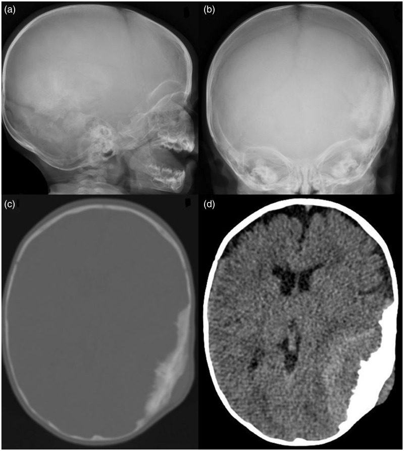
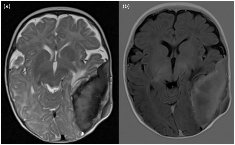
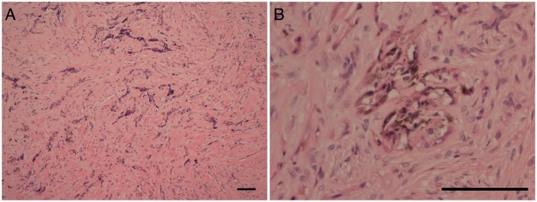
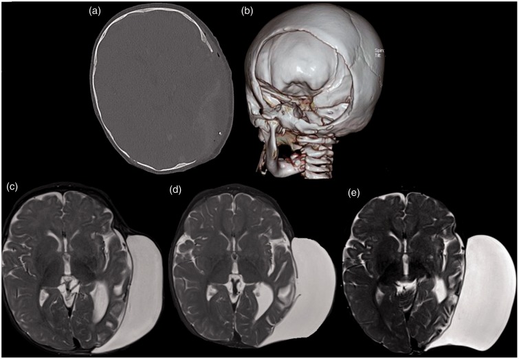
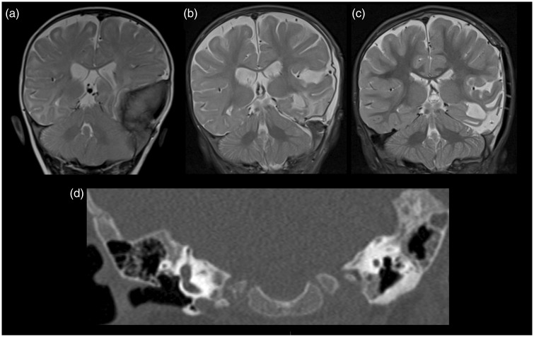
Melanotic neuroectodermal tumour of infancy is an uncommon pigmented neoplasm of neural crest origin. It was first described in 1918 by Krompecher, known as congenital melanocarcinoma at that time. Although it is generally agreed upon that it is a benign entity, it is locally aggressive and has a significant recurrent risk, reported to be between 10-15%. There have also been prior reports of malignant behaviour in these tumours, although extremely rare. The majority of cases of this tumour (about 70%) arise from the maxilla and its occurrence in the cranial vault represents approximately 15.6% of cases. We describe a rare case of melanotic neuroectodermal tumour of infancy, with simultaneous involvement of the cranial vault and petrous temporal bone, in a four-month-old child, complicated by post-surgical pseudo-meningocele. This case illustrates the diagnostic dilemma in differentiating reactive osseous sclerosis from direct tumour infiltration, both of which can occur in the context of melanotic neuroectodermal tumour of infancy. The discussion places emphasis on differential diagnoses and useful radiological features to assist in clinching the diagnosis of melanotic neuroectodermal tumour of infancy.
Keywords: Melanotic neuroectodermal tumour of infancy; cranial vault mass; pigmented neoplasm; pseudo-meningocele.
Figures
Similar articles
-
Melanotic neuroectodermal tumor of infancy arising from the skull: report of an unusual case, review of the literature, and a diagnostic approach.Childs Nerv Syst. 2020 Mar;36(3):469-475. doi: 10.1007/s00381-019-04476-7. Epub 2020 Jan 3. Childs Nerv Syst. 2020. PMID: 31897638 Review.
-
Melanotic neuroectodermal tumor of infancy arising in the temporal bone.J Child Neurol. 2015 Apr;30(5):631-4. doi: 10.1177/0883073813494477. Epub 2013 Aug 20. J Child Neurol. 2015. PMID: 23965399
-
Giant melanotic neuroectodermal tumor of infancy (melanotic progonoma) of the head and neck: report of a malignant case.J Neurosurg Pediatr. 2017 May;19(5):538-545. doi: 10.3171/2016.11.PEDS16509. Epub 2017 Feb 24. J Neurosurg Pediatr. 2017. PMID: 28291424
-
Melanotic neuroectodermal tumor of infancy in the skull: case report and review of the literature.Surg Neurol. 2005 Mar;63(3):275-80. doi: 10.1016/j.surneu.2004.02.032. Surg Neurol. 2005. PMID: 15734527
-
Melanotic neuroectodermal tumor of infancy (MNT1) arising in the skull. Short review of two cases.Acta Neurochir (Wien). 2010 May;152(5):869-75. doi: 10.1007/s00701-009-0472-5. Epub 2009 Aug 11. Acta Neurochir (Wien). 2010. PMID: 19669690 Review.
Cited by
-
Melanotic neuroectodermal tumor of infancy arising from the skull: report of an unusual case, review of the literature, and a diagnostic approach.Childs Nerv Syst. 2020 Mar;36(3):469-475. doi: 10.1007/s00381-019-04476-7. Epub 2020 Jan 3. Childs Nerv Syst. 2020. PMID: 31897638 Review.
-
Radiologic diagnosis of non-traumatic paediatric head and neck emergencies.Pediatr Radiol. 2023 Apr;53(4):768-782. doi: 10.1007/s00247-022-05556-8. Epub 2022 Dec 9. Pediatr Radiol. 2023. PMID: 36481939 Review.
References
-
- Borello ED, Gorlin RJ. Melanotic neuroectodermal tumor of infancy–a neoplasm of neural crest origin. Report of a case associated with high urinary excretion of vanilmandelic acid. Cancer 1966; 19: 196–206. - PubMed
-
- Cutler LS, Chaudhry AP, Topazian R. Melanotic neuroectodermal tumor of infancy: An ultrastructural study, literature review, and reevaluation. Cancer 1981; 48: 257–270. - PubMed
-
- Zimmerman RA, Bilaniuk LT. CT of primary and secondary craniocerebral neuroblastoma. AJR Am J Roentgenol 1980; 135: 1239–1242. - PubMed
-
- Rachidi S, Sood AJ, Patel KG, et al. Melanotic neuroectodermal tumor of infancy: A systematic review. J Oral Maxillofac Surg 2015; 73: 1946–1956. - PubMed
Publication types
MeSH terms
LinkOut - more resources
Full Text Sources
Other Literature Sources
