

Detection accuracy for epithelial dysplasia using an objective autofluorescence visualization method based on the luminance ratio
- PMID: 29125138
- PMCID: PMC5775331
- DOI: 10.1038/ijos.2017.37
Detection accuracy for epithelial dysplasia using an objective autofluorescence visualization method based on the luminance ratio
Abstract
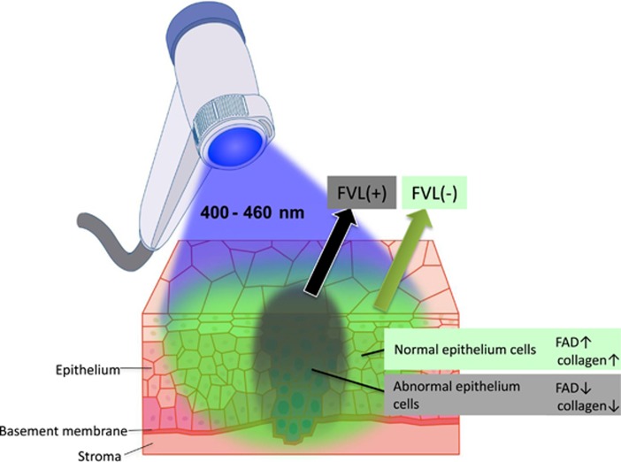
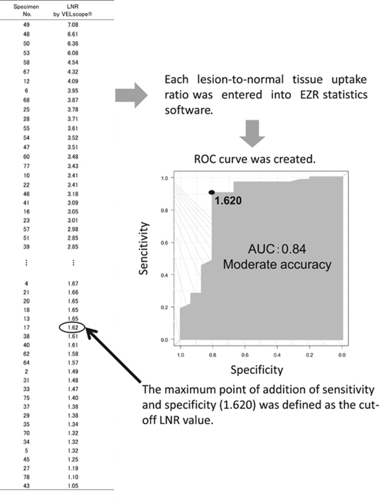
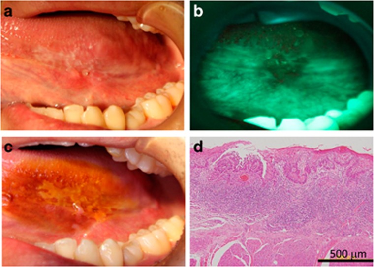
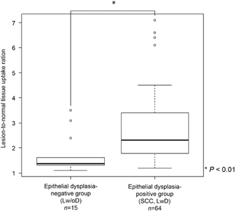
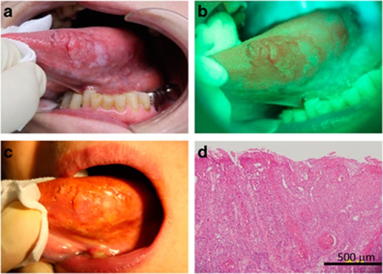
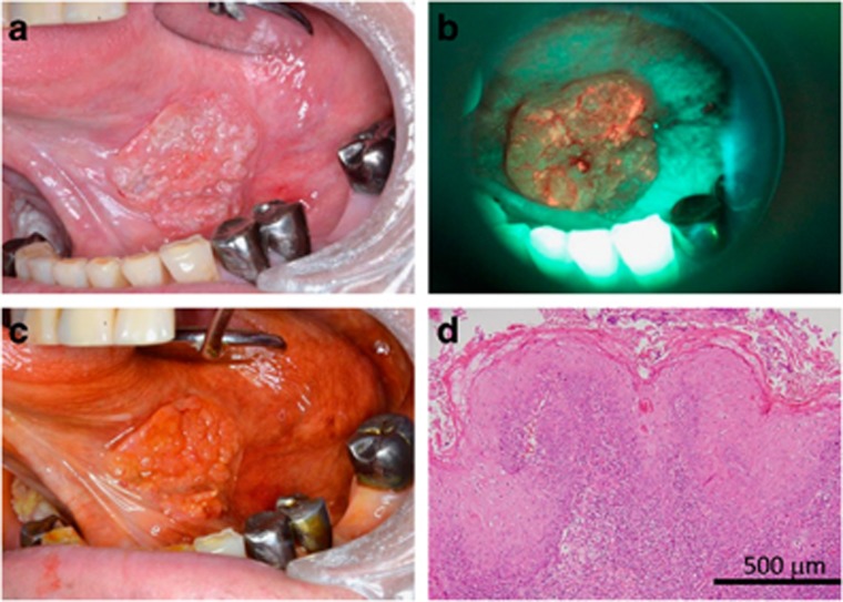
The autofluorescence visualization method (AVM) uses blue excitation light to assist in the diagnosis of epithelial dysplasia. It detects epithelial dysplasia as a black area, which is known as fluorescence visualization loss (FVL). In this study, we evaluated the detection accuracy for epithelial dysplasia of the tongue using the objective AVM and assessed its possible clinical utility. Seventy-nine tongue specimens clinically suspected to have leukoplakia or squamous cell carcinoma (SCC) were analyzed. First, the AVM was subjectively performed using the Visually Enhanced Lesion scope (VELscope), and the iodine-staining method was then performed. After biopsy, the histopathological results and the luminance ratio between the lesion and healthy tissue were compared, and a receiver operating characteristic curve was created. The cutoff value for the objective AVM was determined; the lesion was considered FVL-positive or -negative when the luminance ratio was higher or lower than the cutoff value, respectively. The histopathological diagnoses among the 79 specimens were SCC (n=30), leukoplakia with dysplasia (n=34), and leukoplakia without dysplasia (n=15). The cutoff value of the luminance ratio was 1.62, resulting in 66 FVL-positive and 13 FVL-negative specimens. The luminance ratio was significantly higher in the epithelial dysplasia-positive than -negative group (P<0.000 1). The objective AVM showed much higher consistency between histopathological results than did the two methods (kappa statistic=0.656). In conclusion, objective autofluorescence visualization has a potential as an auxiliary method for diagnosis of epithelial dysplasia.
Figures
References
-
- Kuribayashi Y, Tsushima F, Sato M et al. Recurrence patterns of oral leukoplakia after curative surgical resection: important factors that predict the risk of recurrence and malignancy. J Oral Pathol Med 2012; 41 (9): 682–688. - PubMed
-
- Arnaoutakis D, Bishop J, Westra W et al. Recurrence patterns and management of oral cavity premalignant lesions. Oral Oncol 2013; 49 (8): 814–817. - PubMed
-
- Kuribayashi Y, Tsushima F, Morita K et al. Long-term outcome of non-surgical treatment in patients with oral leukoplakia. Oral Oncol 2015; 51 (11): 1020–1025. - PubMed
-
- Yajima Y, Noma H, Furuya Y et al. Quantification of telomerase activity of regions unstained with iodine solution that surround oral squamous cell carcinoma. Oral Oncol 2004; 40 (3): 314–320. - PubMed
-
- Banoczy J, Sugar L. Longitudinal studies in oral leukoplakias. J Oral Pathol 1972; 1 (6): 265–272. - PubMed
MeSH terms
LinkOut - more resources
Full Text Sources
Other Literature Sources
Medical
Research Materials
