

Advanced skeletal maturity in children and adolescents with myelomeningocele
- PMID: 29125519
- PMCID: PMC6373769
- DOI: 10.3233/PRM-170458
Advanced skeletal maturity in children and adolescents with myelomeningocele
Abstract
Purpose: Atypical skeletal development is common in youth with myelomeningocele (MM), though the underlying reasons have not been fully elucidated. This study assessed skeletal maturity in children and adolescents with MM and examined the effects of sex, age, sexual development, ethnicity, anthropometrics and shunt status.
Methods: Forty-three males and 35 females with MM, 6-16 years old, underwent hand radiographs for bone age determination. The difference between bone age and chronological age was evaluated using Wilcoxon sign rank tests. Relationships between age discrepancy (skeletal-chronological) and participant characteristics were assessed using multiple linear regression with forward selection.
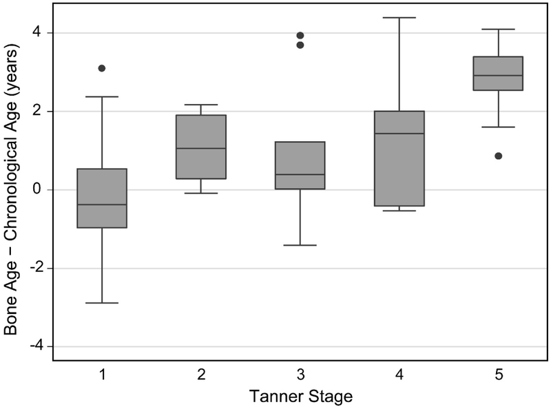
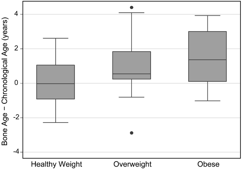
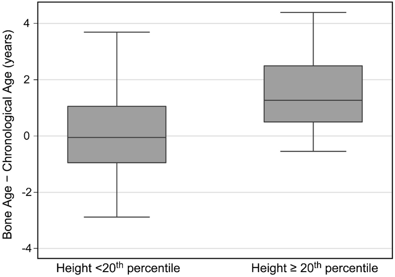
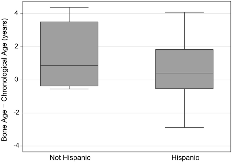
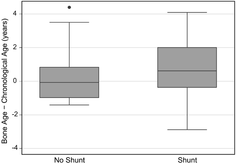
Results: Overall, forty percent (31/78) of MM participants had an advanced bone age of 1 year or greater (median: 2.5 years), while 47% (37/78) were within 1 year above or below their chronological age (-0.001 years) and 13% (10/78) were delayed by more than 1 year (-1.4 years). Bone age was advanced compared to chronologic age in both males and females (p⩽ 0.024). Advanced bone age was observed in early to late puberty and after maturation (p⩽ 0.07), as well as in Hispanic participants (p= 0.003) and in those with a shunt (p= 0.0004). Advanced bone age was positively correlated with height, weight and body mass index (BMI) percentiles (p= 0.004). In multiple linear regression analysis, advanced bone age was most strongly associated with higher Tanner stage of sexual development, and higher weight, height or BMI percentile.
Conclusions: Advanced skeletal maturity is common in children/adolescents with MM over 8 years of age who have reached puberty (65%), particularly those who are overweight (80%). Hormonal effects associated with adiposity and sexual maturity likely influence skeletal maturation. Clinicians may use Tanner stage and weight or BMI to gain insight into skeletal maturity.
Keywords: Skeletal maturity; bone age; myelomeningocele; pediatrics; spina bifida.
Conflict of interest statement
Figures
Similar articles
-
The impact of HIV infection on skeletal maturity in peripubertal children in Zimbabwe: a cross-sectional study.BMC Pediatr. 2024 Jul 27;24(1):480. doi: 10.1186/s12887-024-04965-y. BMC Pediatr. 2024. PMID: 39068422 Free PMC article.
-
Advanced bone age in children with Blount disease: a case-control study.J Pediatr Orthop. 2013 Jul-Aug;33(5):551-7. doi: 10.1097/BPO.0b013e318285c524. J Pediatr Orthop. 2013. PMID: 23752155
-
Skeletal maturity in myelomeningocele.J Pediatr Orthop. 2003 Nov-Dec;23(6):718-21. doi: 10.1097/00004694-200311000-00007. J Pediatr Orthop. 2003. PMID: 14581773
-
Skeletal age assessment from elbow radiographs. Review of the literature.Chir Organi Mov. 2008 May;92(1):1-6. doi: 10.1007/s12306-008-0032-9. Epub 2008 Apr 11. Chir Organi Mov. 2008. PMID: 18408902 Review.
-
Using Skeletal Maturity in Pediatric Orthopaedics: A Primer.J Pediatr Orthop. 2022 Aug 1;42(7):e793-e800. doi: 10.1097/BPO.0000000000002107. Epub 2022 Mar 23. J Pediatr Orthop. 2022. PMID: 35316260 Review.
Cited by
-
Quantitative Computed Tomography Assessment of Bone Deficits in Ambulatory Children and Adolescents with Spina Bifida: Importance of Puberty.JBMR Plus. 2020 Nov 30;4(12):e10427. doi: 10.1002/jbm4.10427. eCollection 2020 Dec. JBMR Plus. 2020. PMID: 33354646 Free PMC article.
-
Nutrition, metabolic syndrome, and obesity: Guidelines for the care of people with spina bifida.J Pediatr Rehabil Med. 2020;13(4):637-653. doi: 10.3233/PRM-200753. J Pediatr Rehabil Med. 2020. PMID: 33325412 Free PMC article. Review.
References
-
- Bowman RM, Boshnjaku V, McLone DG. The changing incidence of myelomeningocele and its impact on pediatric neurosurgery: a review from the Children’s Memorial Hospital. Child’s nervous system : ChNS : official journal of the International Society for Pediatric Neurosurgery. 2009. July;25(7):801–6. PubMed PMID: 19326126. Epub 2009/03/28. eng. - PubMed
-
- Apkon SD, Fenton L, Coll JR. Bone mineral density in children with myelomeningocele. Developmental medicine and child neurology. 2009. January;51(1):63–7. PubMed PMID: 18811711. Epub 2008/09/25. eng. - PubMed
-
- Ausili E, Focarelli B, Tabacco F, Fortunelli G, Caradonna P, Massimi L, et al. Bone mineral density and body composition in a myelomeningocele children population: effects of walking ability and sport activity. European review for medical and pharmacological sciences. 2008. Nov-Dec;12(6):349–54. PubMed PMID: 19146196. Epub 2009/01/17. eng. - PubMed
-
- Quan A, Adams R, Ekmark E, Baum M. Bone mineral density in children with myelomeningocele. Pediatrics. 1998. September;102(3):E34. PubMed PMID: 9724682. Epub 1998/09/02. eng. - PubMed
-
- Feeley BT, Ip TC, Otsuka NY. Skeletal maturity in myelomeningocele. Journal of pediatric orthopedics. 2003. Nov-Dec;23(6):718–21. PubMed PMID: 14581773. Epub 2003/10/29. eng. - PubMed
Publication types
MeSH terms
Grants and funding
LinkOut - more resources
Full Text Sources
Other Literature Sources
