Review
. 2018 Jan 24:13:351-378.
doi: 10.1146/annurev-pathol-020117-044010. Epub 2017 Nov 10.

Wnt/β-Catenin Signaling in Liver Development, Homeostasis, and Pathobiology

Affiliations
Review

Wnt/β-Catenin Signaling in Liver Development, Homeostasis, and Pathobiology

Jacquelyn O Russell et al. Annu Rev Pathol. .

Abstract

The liver is an organ that performs a multitude of functions, and its health is pertinent and indispensable to survival. Thus, the cellular and molecular machinery driving hepatic functions is of utmost relevance. The Wnt signaling pathway is one such signaling cascade that enables hepatic homeostasis and contributes to unique hepatic attributes such as metabolic zonation and regeneration. The Wnt/β-catenin pathway plays a role in almost every facet of liver biology. Furthermore, its aberrant activation is also a hallmark of various hepatic pathologies. In addition to its signaling function, β-catenin also plays a role at adherens junctions. Wnt/β-catenin signaling also influences the function of many different cell types. Due to this myriad of functions, Wnt/β-catenin signaling is complex, context-dependent, and highly regulated. In this review, we discuss the Wnt/β-catenin signaling pathway, its role in cell-cell adhesion and liver function, and the cell type-specific roles of Wnt/β-catenin signaling as it relates to liver physiology and pathobiology.

Keywords: cholangiocyte; hepatoblastoma; hepatocellular cancer; hepatocyte; liver stem cell; liver tumors; regeneration; zonation.


Figures

Figure 1
Figure 1
The canonical Wnt signaling pathway. (a) In the absence of Wnt binding to its receptor (Frizzled) and coreceptor (LRP5/6), β-catenin is phosphorylated by its destruction complex and targeted for proteasomal degradation by βTRCP. The Frizzled receptor is targeted for proteasomal degradation via the activity of ZNRF3/RNF43. (b) Upon release of biologically active Wnt from a neighboring cell by cargo receptor Wntless, the Wnt protein binds its receptor and coreceptor, which triggers recruitment of the β-catenin destruction complex to the plasma membrane through scaffolding protein Dishevelled. This interaction is further stabilized by R-spondin binding to an LGR receptor. β-catenin cannot be phosphorylated, accumulates in the cytoplasm, and translocates to the nucleus to bind the TCF/LEF family of transcription factors to induce target gene transcription. Abbreviations: APC, adenomatous polyposis coli; β-cat, β-catenin; βTRCP, β-transducin repeat-containing protein; CK1α, casein kinase 1α; Dvl, Dishevelled; GSK3β, glycogen synthase kinase 3β; LGR, leucine-rich repeat-containing G protein–coupled receptor; LRP, lipoprotein receptor-related protein; RNF43, ring finger 43; TCF/LEF, T cell factor/lymphoid enhancer factor; ZNRF3, zinc and ring finger 3.
Figure 2
Figure 2
Noncanonical Wnt signaling pathway. (Left) In the planar cell polarity pathway, Wnt ligands bind to a complex consisting of certain Frizzled receptors, Ror2, and Dishevelled, which triggers activation of RhoA and ROCK or, alternatively, activation of Rac and JNK signaling, to regulate cell polarity and migration. (Right) In the Wnt/calcium pathway, Wnt ligands bind to a complex consisting of Frizzled receptors, Dishevelled, and G proteins, which leads to the activation of PLC and the generation of DAG and IP3. DAG activates PKC whereas IP3 promotes increased intracellular calcium levels, which leads to the activation of CaMKII and CaN, which, in turn, regulates cell migration and proliferation. Abbreviations: βγ, G protein signaling subunit β/γ; CaMKII, calcium/calmodulin-dependent; CaN, calcineurin; DAG, diacylglycerol; Dvl, Dishevelled; Gα, G protein subunit α; IP3, inositol 1,4,5-triphosphate; JNK, c-Jun N-terminal kinase; PCP, planar cell polarity; PKC, protein kinase C; PLC, phospholipase C; PIP2, phosphatidylinositol 4,5 biphosphate; Rac, Ras-related C3 botulinum toxin substrate; RhoA, Ras homolog gene family, member A; ROCK, Rho-associated protein kinase; Ror2, receptor tyrosine kinase-like orphan receptor 2.
Figure 3
Figure 3
Wnt/TOR and Wnt/STOP signaling pathways. (Left) In the Wnt/TOR pathway, in the absence of Wnt ligands, GSK3β phosphorylates and activates TSC2, which, in turn, inhibits mTORC1 activity. Binding of the Wnt protein to its receptor and coreceptor leads to sequestration of the destruction complex, including GSK3β, into multivesicular bodies. This prevents activation of TSC2, leading to activation of mTORC1 and promotion of protein translation. (Right) In the Wnt/STOP pathway, the activity of GSK3β promotes phosphorylation and proteasomal degradation of a multitude of target proteins. Upon Wnt ligand binding and sequestration of GSK3β into multivesicular bodies, these GSK3β-target proteins are no longer targeted for degradation and accumulate in the cytoplasm. Abbreviations: APC, adenomatous polyposis coli; CK1α, casein kinase 1α; Dvl, Dishevelled; GSK3β, glycogen synthase kinase 3β; LRP, lipoprotein receptor-related protein; mTOR, mechanistic target of rapamycin; mTORC1, mTOR complex 1; TSC2, tuberous sclerosis complex 2; Wnt/STOP, Wnt-dependent stabilization of proteins; Wnt/TOR, Wnt-dependent regulation of mTOR.
Figure 4
Figure 4
Architecture of the hepatic sinusoid and metabolic zonation. The hepatic sinusoid is composed of multiple, carefully organized cell types. The main epithelial cells of the liver, the hepatocytes, are arranged in chords stretching from a portal triad, which consists of the portal vein, bile duct, and hepatic artery, to the central vein. These chords of hepatocytes are lined by specialized LSECs, which allow optimal perfusion of hepatocytes by the sinusoidal blood. Hepatic stellate cells reside in the space between hepatocytes and LSECs, whereas the tissue-resident macrophages, known as Kupffer cells, are adherent to LSECs within the hepatic sinusoid. The second epithelial cell type of the liver, biliary epithelial cells, lines the bile ducts in the portal triad. In terms of metabolic zonation, the hepatocytes adjacent to the portal triad constitute zone 1, where HNF4α binds to TCF to promote expression of the periportal gene signature. This zone is also characterized by periportal YAP signaling, which decreases in a gradient with increasing distance from the portal triad. The hepatocytes lining the central vein constitute zone 3, characterized by active Wnt/β-catenin driven via Wnt2 and Wnt9b secretion from venous endothelial cells. In this zone, β-catenin binds to TCF to promote expression of the pericentral gene signature. The hepatocytes in between zones 1 and 3 constitute zone 2, and display relatively low YAP and Wnt/β-catenin signaling. Abbreviations: β-cat, β-catenin; HNF4α, hepatocyte nuclear factor 4α; LSECs, liver sinusoidal endothelial cells; TCF, T cell factor; YAP, Yes-associated protein.
Figure 5
Figure 5
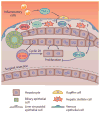
Role of Wnt/β-catenin signaling in liver regeneration following surgical resection. Following a surgical resection of liver mass, Wnt/β-catenin signaling is activated to promote liver regeneration. Infiltrating inflammatory cells secrete TNFα, which promotes Wnt expression in macrophages. Additionally, liver sinusoidal endothelial cells secrete Wnt2 and HGF; the latter is also secreted by hepatic stellate cells. Neither biliary epithelial cells nor hepatocytes secrete mitogenic Wnts following surgical resection. The secreted Wnt ligands act on hepatocytes to promote β-catenin translocation to the nucleus, where it promotes expression of target genes, such as cyclin D1, to promote hepatocyte proliferation. Following the restoration of sufficient liver mass, hepatocytes secrete Wnt5a to inhibit canonical Wnt/β-catenin signaling and promote termination of liver regeneration. Abbreviations: β-cat, β-catenin; HGF, hepatocyte growth factor; TNFα, tumor necrosis factor α.
Figure 6
Figure 6
Role of β-catenin in liver tumors. (a) Oncogenic activation of β-catenin occurs in multiple types of liver cancer, including HCC and HB. Common mechanisms of β-catenin activation include missense mutations and exon-3 deletions. In HCC, oncogenic β-catenin activation cooperates with mutations in proteins such as ARID2, NFE2L2, TERT, APOB, and MLL2 or with Met activation to drive tumorigenesis. Interestingly, mutations in TP53 and AXIN1 or YAP activation tend to be mutually exclusive with β-catenin activation in HCC. In contrast, concomitant activation of β-catenin and YAP signaling is observed in approximately 80% of HB tumor samples, which suggests there are distinct mechanisms of hepatocarcinogenesis involving β-catenin and YAP in HB and HCC. (b) Wnt/β-catenin signaling activation regulates many aspects of tumor biology, as various downstream targets promote multiple oncogenic processes, including proliferation, survival, metabolism, immune tolerance, and angiogenesis. Abbreviations: EpCAM, epithelial cell adhesion molecule; HB, hepatoblastoma; HCC, hepatocellular carcinoma; Lect2, leukocyte cell–derived chemotaxin 2; Lgr5, leucine-rich repeat-containing G protein–coupled receptor 5; VEGF-A, vascular endothelial growth factor A; YAP, Yes-associated protein.
Figure 7
Figure 7
Role of Wnt/β-catenin signaling in stem cell–mediated liver regeneration. (a) In conditions of extreme liver injury associated with a block of hepatocyte proliferation, bipotent liver progenitor cells (LPCs) arise from the biliary epithelial cell (BEC) compartment. Secretion of Wnt3a from Kupffer cells induces Wnt/β-catenin signaling in LPCs and promotes their transdifferentiation into hepatocytes to mediate regeneration from hepatocyte injury. Alternatively, induction of Notch signaling through Jagged1 promotes LPC transdifferentiation to BECs to mediate regeneration from biliary injury. (b) During conditions of biliary injury, such as that induced by a DDC diet, BECs secrete Wnt7a, which induces Sox9 expression and β-catenin activation in hepatocytes and promotes their transdifferentiation into BECs. Additionally, BECs secrete Wnt7b and Wnt10a to promote proliferation of BECs in a β-catenin-independent manner. Abbreviations: BEC, biliary epithelial cell; β-cat, β-catenin; DDC, hepatotoxin 3,5-diethoxycarbonyl-1, 4-dihydrocollidine; LPC, liver progenitor cell.

References

    1. Kimelman D, Xu W. β-catenin destruction complex: insights and questions from a structural perspective. Oncogene. 2006;25:7482–91. - PubMed
    1. Behrens J, Jerchow BA, Würtele M, Grimm J, Asbrand C, et al. Functional interaction of an axin homolog, conductin, with β-catenin, APC, and GSK3β. Science. 1998;280:596–99. - PubMed
    1. Liu C, Li Y, Semenov M, Han C, Baeg GH, et al. Control of β-catenin phosphorylation/degradation by a dual-kinase mechanism. Cell. 2002;108:837–47. - PubMed
    1. Amit S, Hatzubai A, Birman Y, Andersen JS, Ben-Shushan E, et al. Axin-mediated CKI phosphorylation of β-catenin at Ser 45: a molecular switch for the Wnt pathway. Genes Dev. 2002;16:1066–76. - PMC - PubMed
    1. Aberle H, Bauer A, Stappert J, Kispert A, Kemler R. β-catenin is a target for the ubiquitin-proteasome pathway. EMBO J. 1997;16:3797–804. - PMC - PubMed

Publication types