

Nitrergic neurons of the dorsal raphe nucleus encode information about stress duration
- PMID: 29125838
- PMCID: PMC5681257
- DOI: 10.1371/journal.pone.0187071
Nitrergic neurons of the dorsal raphe nucleus encode information about stress duration
Abstract
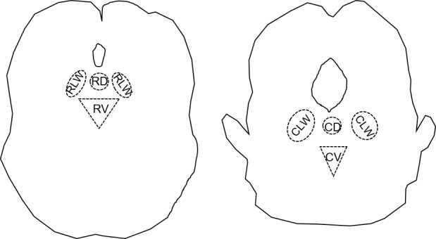
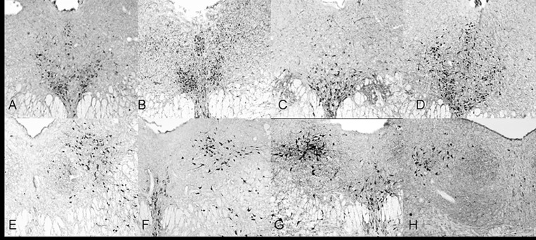
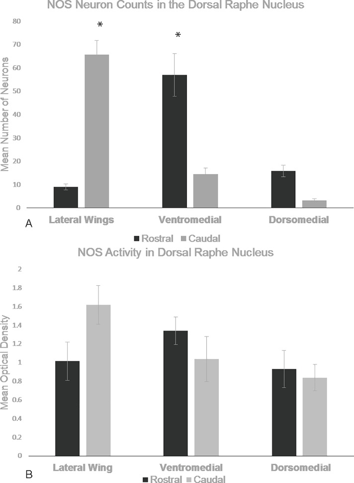
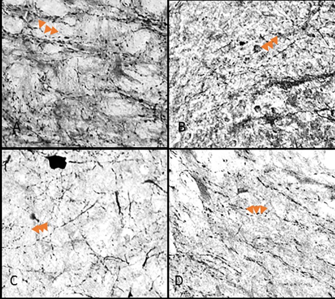
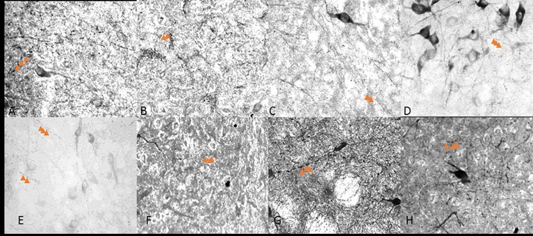
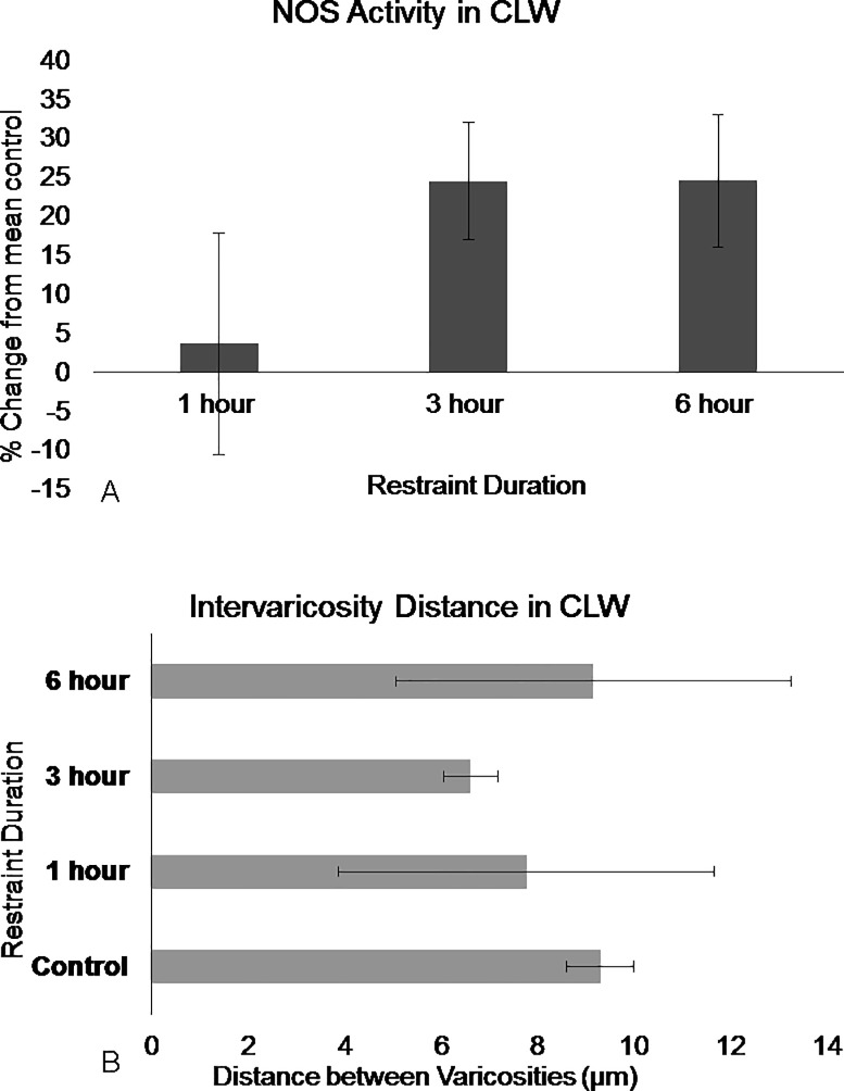
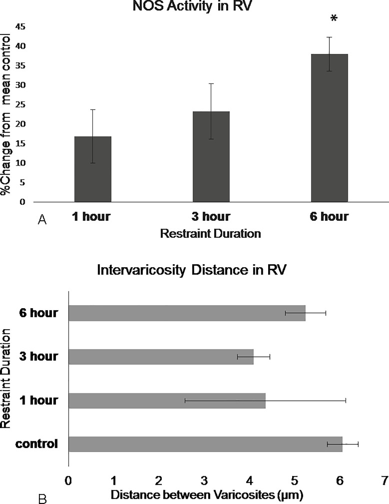
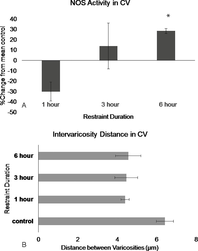
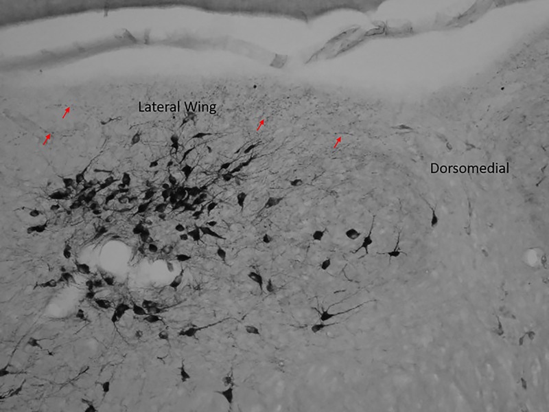
Nitrergic neurons of the dorsal raphe nucleus (DRN) may play a role in physiological stress responses. The caudal lateral wings (CLW) are unique compared to other rostral-caudal DRN sub-regions because they contain distinct nitric oxide (NO) synthase (NOS) populations that are independent of tryptophan hydroxylase (TPH). NOS neurons in the CLW are also highly activated during acute restraint stress. However, the effects of acute stress duration on NOS activation in the CLW are unclear. Here NADPH-d, an index of NOS activity, is used to show that sub-regions of the DRN have differential NOS activation in response to 6 hours of restraint stress in rats. We report increased NOS activity through 6 hours of restraint in the caudal lateral wings and ventromedial sub-regions. These data suggest that, NOS neurons may play a dynamic role in the response to stress duration.
Conflict of interest statement
Figures
Similar articles
-
Acute restraint increases NADPH-diaphorase staining in distinct subregions of the rat dorsal raphe nucleus: implications for raphe serotonergic and nitrergic transmission.Brain Res. 2006 Nov 13;1119(1):174-81. doi: 10.1016/j.brainres.2006.08.058. Epub 2006 Sep 20. Brain Res. 2006. PMID: 16989783
-
Cellular profile of the dorsal raphe lateral wing sub-region: relationship to the lateral dorsal tegmental nucleus.J Chem Neuroanat. 2014 May;57-58:15-23. doi: 10.1016/j.jchemneu.2014.03.001. Epub 2014 Apr 3. J Chem Neuroanat. 2014. PMID: 24704911 Free PMC article.
-
Activity-dependent heterogeneous populations of nitric oxide synthase neurons in the rat dorsal raphe nucleus.Brain Res. 2006 May 1;1086(1):117-32. doi: 10.1016/j.brainres.2006.02.107. Epub 2006 Apr 17. Brain Res. 2006. PMID: 16616732
-
Functional organization of the dorsal raphe efferent system with special consideration of nitrergic cell groups.J Chem Neuroanat. 2011 Jul;41(4):281-93. doi: 10.1016/j.jchemneu.2011.05.008. Epub 2011 May 24. J Chem Neuroanat. 2011. PMID: 21640185 Review.
-
GABAergic modulation of serotonergic neurons in the dorsal raphe nucleus.Rev Neurosci. 2019 Apr 24;30(3):289-303. doi: 10.1515/revneuro-2018-0014. Rev Neurosci. 2019. PMID: 30173207 Review.
Cited by
-
GABAergic interneurons' feedback inhibition of dorsal raphe-projecting pyramidal neurons of the medial prefrontal cortex suppresses feeding of adolescent female mice undergoing activity-based anorexia.Brain Struct Funct. 2022 Jul;227(6):2127-2151. doi: 10.1007/s00429-022-02507-9. Epub 2022 May 30. Brain Struct Funct. 2022. PMID: 35635653 Free PMC article.
-
Selective vulnerability of dorsal raphe-medial prefrontal cortex projection neurons to corticosterone-induced hypofunction.Eur J Neurosci. 2019 Jul;50(1):1712-1726. doi: 10.1111/ejn.14355. Epub 2019 Feb 15. Eur J Neurosci. 2019. PMID: 30687960 Free PMC article.
-
Evidence for chronic headaches induced by pathological changes of myodural bridge complex.Sci Rep. 2024 Mar 4;14(1):5285. doi: 10.1038/s41598-024-55069-7. Sci Rep. 2024. PMID: 38438423 Free PMC article.
-
Sleep loss mediates the effect of stress on nitrergic signaling in female mice.Neurosci Lett. 2021 Jan 1;740:135362. doi: 10.1016/j.neulet.2020.135362. Epub 2020 Nov 6. Neurosci Lett. 2021. PMID: 33166635 Free PMC article.
References
-
- Xu ZQ, Hokfelt T. Expression of galanin and nitric oxide synthase in subpopulations of serotonin neurons of the rat dorsal raphe nucleus. J Chem Neuroanat, 1997. 13(3): p. 169–87. - PubMed
-
- Vasudeva RK, Lin RC, Simpson KL, Waterhouse BD. Functional organization of the dorsal raphe efferent system with special consideration of nitrergic cell groups. J Chem Neuroanat, 2011. 41(4): p. 281–93. doi: 10.1016/j.jchemneu.2011.05.008 - DOI - PubMed
-
- Okere CO, Waterhouse BD. Activity-dependent heterogeneous populations of nitric oxide synthase neurons in the rat dorsal raphe nucleus. Brain Res, 2006. 1086. - PubMed
-
- Spiacci A, Coimbra NC, Zangrossi H. Differential Involvement of Dorsal Raphe Subnuclei in the regulation of anxiety- and panic- related defensive behaviors. Neuroscience, 2012. 227: p 350–360 doi: 10.1016/j.neuroscience.2012.09.061 - DOI - PubMed
MeSH terms
Substances
Grants and funding
LinkOut - more resources
Full Text Sources
Other Literature Sources
