. 2017 Nov 10;13(11):e1006727.
doi: 10.1371/journal.ppat.1006727. eCollection 2017 Nov.

Autophagy pathway induced by a plant virus facilitates viral spread and transmission by its insect vector

Affiliations

Autophagy pathway induced by a plant virus facilitates viral spread and transmission by its insect vector

Yong Chen et al. PLoS Pathog. .

Abstract

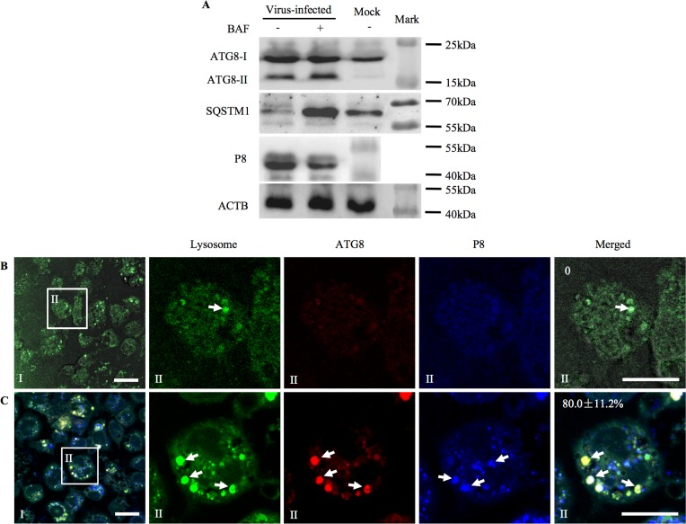
Many viral pathogens are persistently transmitted by insect vectors and cause agricultural or health problems. Generally, an insect vector can use autophagy as an intrinsic antiviral defense mechanism against viral infection. Whether viruses can evolve to exploit autophagy to promote their transmission by insect vectors is still unknown. Here, we show that the autophagic process is triggered by the persistent replication of a plant reovirus, rice gall dwarf virus (RGDV) in cultured leafhopper vector cells and in intact insects, as demonstrated by the appearance of obvious virus-containing double-membrane autophagosomes, conversion of ATG8-I to ATG8-II and increased level of autophagic flux. Such virus-containing autophagosomes seem able to mediate nonlytic viral release from cultured cells or facilitate viral spread in the leafhopper intestine. Applying the autophagy inhibitor 3-methyladenine or silencing the expression of Atg5 significantly decrease viral spread in vitro and in vivo, whereas applying the autophagy inducer rapamycin or silencing the expression of Torc1 facilitate such viral spread. Furthermore, we find that activation of autophagy facilitates efficient viral transmission, whereas inhibiting autophagy blocks viral transmission by its insect vector. Together, these results indicate a plant virus can induce the formation of autophagosomes for carrying virions, thus facilitating viral spread and transmission by its insect vector. We believe that such a role for virus-induced autophagy is common for vector-borne persistent viruses during their transmission by insect vectors.


Conflict of interest statement

The authors have declared that no competing interests exist.

Figures

Fig 1
Fig 1. RGDV infection activated the autophagy pathway in VCMs of R. dorsalis.
(A, B) At 48 hpi, ATG8 in mock- or virus-infected VCMs treated with (+) and without (-) 3-MA or BFA was detected by western blot assay using ATG8-specifc IgG. Insect ACTB was detected with ACTB-specific IgG as a control. (C-E) Autophagosomes in mock (D) and virus-infected (E) VCMs were detected by immunofluorescence microscopy. At 48 hpi, VCMs were immunolabeled for autophagosomes with ATG8-FITC (green), for viral particles with P8-rhodamine (red), and for viroplasms with Pns9-Alexa Fluor 647 (blue), and then processed for immunofluorescence microscopy. Viral antigens surrounding the spherical structures composed of Pns9, a component of viroplasms, are indicated by arrowheads. Arrows indicate the colocalization of ATG8-specific autophagosomes and viral particles. The average number of ATG8-specific puncta per R. dorsalis cell and a minimum of 100 cells were counted (C), and data points presented are the averages from six different fields (D, E). *P < 0.05. Bars, 15 μm. (F-L) Transmission electron micrographs for virus-induced autophagosomes in VCMs. Representative images were shown for mock VCMs (F), and for virus-containing autophagosomes in the cytoplasm (G) or at the periphery of viroplasm at 48 hpi (H). Panel II is an enlargement of the boxed area in panel I. Red arrows in panel G indicate double-membrane of autophagosomes. Red arrow in panel H indicates single-membrane of autophagosomes. (I) Virus-infected VCMs at 48 hpi were immunolabeled with ATG8-specific IgG as the primary antibody, followed by treatment with 10-nm gold particle-conjugated goat antibodies against rabbit IgG as secondary antibodies. Panel II is an enlargement of the boxed area in panel I. Arrows indicate gold particles. (J-L) At 48 hpi, virus-containing autophagosomes inside (J), at the periphery (K), or outside (L) of infected cells. (M) The quantification of the average number of autophagosome-like vesicles per R. dorsalis cell and a minimum of 30 cells were counted. *P < 0.05. M, mitochondrion. N, nucleus. Vi, virion. VP, viroplasm. Bars in panel F (I) and (II), 500 nm. Bars in panels G-L, 200 nm.
Fig 2
Fig 2. RGDV infection increased the level of autophagic flux in VCMs of R. dorsalis.
(A) At 48 hpi, RGDV P8, ATG8 and SQSTM1 in VCMs treated with (+) and without (-) BAF were detected by western blot assay. Insect ACTB was detected with ACTB-specific IgG as a control. (B, C) Fusion of virus-induced autophagosomes with lysosomes in mock- (B) or virus-infected (C) VCMs, as revealed by immunofluorescence microscopy. At 48 hpi, VCMs were immunolabeled for lysosomes with LysoTracker Green DND-26 (green), for autophagosomes with ATG8-rhodamine (red), and for viral particles with P8-Alexa Fluor 647 (blue), and then processed for confocal microscopy. Data points presented are the averages from six different fields (B, C). Bars, 15 μm.
Fig 3
Fig 3. Autophagy pathway induced by RGDV infection facilitated viral nonlytic release from VCMs of R. dorsalis.
(A) Percentage of insect vector cells infected with RGDV. The VCMs were transfected for 8 h with rapamycin or 3-MA (panels I) or dsAtg5 or dsTorc1 (panels II), then inoculated with RGDV at a MOI of 0.1 for 2 h. At 48 hpi, cells were immunolabeled with ATG8-FITC (green) and P8-rhodamine (red), then examined with confocal microscopy. Arrows indicate the colocalization of ATG8-specific autophagosomes and viral particles. Data points presented are the averages from six different fields (A). Bars, 20 μm. (B) At 48 and 72 hpi, effects of rapamycin or 3-MA (panels I) and dsAtg5 or dsTorc1 (panels II) on transcript levels of RGDV P8 gene in VCMs as revealed by RT-qPCR assay. Means (±standard deviation [SD]) from three biological replicates are shown. *P < 0.05. (C) At 48 hpi, effects of rapamycin or 3-MA (panels I) and dsAtg5 or dsTorc1 (panels II) on transcript levels of Atg8 gene in VCMs as revealed by RT-qPCR assay. Means (±SD) from three biological replicates are shown. *P < 0.05. (D) The accumulation levels of ATG8 and SQSTM1 were detected by western blot assay using ATG8- and SQSTM1-specifc IgG, respectively. Insect ACTB was detected with ACTB-specific IgG as a control. (E) Autophagy induced by viral infection increased the extracellular viral RNA levels. VCMs were transfected for 8 h with dsRNAs, then inoculated with RGDV at a MOI of 10 for 2 h. At 48 and 72 hpi, culture supernatant was collected to measure the viral titers detected by RT-qPCR assay. Means (±SD) from three biological replicates are shown. *P < 0.05.
Fig 4
Fig 4. RGDV infection triggered autophagy pathway in the intestine of R. dorsalis.
(A) At 4 days padp, ATG8 and SQSTM1 in nonviruliferous or viruliferous insects were detected by western blot assay using ATG8- and SQSTM1-specifc IgG, respectively. Insect ACTB was detected with ACTB-specific IgG as a control. ATG8-I was detected in both nonvirulifeorus and viruliferous insects, and ATG8-II was only detected in viruliferous insects. (B-D) Autophagosomes in nonviruliferous (B) or viruliferous (C, D) insects were detected by confocal microscopy. At 4 days padp, insect intestines were immunolabeled for autophagosomes with ATG8-FITC (green), for viral particles with P8-rhodamine (red), and for ACTB with ACTB dye phalloidin-Alexa Fluor 647 (blue), then examined with confocal microscopy. Panels II are enlargements of boxed areas in panels I. Single optical sections of epithelial side (C) and muscle side (D) of the intestines from viruliferous R. dorsalis are shown from a confocal Z-stack at 100 μm depth. Red arrows indicate colocalization of ATG8-specific autophagosomes and P8-specific viral particles. Bars, 30 μm. (E-J) RGDV infection induced autophagosome formation as measured by electron microscopy. (E) Representative images were shown for nonviruliferous intestinal epithelium. Panel II was an enlargement of the boxed area in panel I. (F) Viruliferous intestinal epithelium was immunolabeled with ATG8-specific IgG as the primary antibody, followed by treatment with 10-nm gold particle-conjugated goat antibodies against rabbit IgG as secondary antibodies. Red arrows indicate gold particles. (G-J) Transmission electron micrographs of virus-containing autophagosomes within the epithelial cytoplasm (G), along the microvilli (H), in the gut lumen (I), or in the visceral muscles tissues (J) of insect intestines. Inset in panel H was the enlargement of the boxed area. Red arrow indicated the double-membrane of virus-containing autophagosome. Vi, virion. CM, circular muscle. LM, longitudinal muscle. BL, basal lamina. Mv, microvilli. M, mitochondrion. N, nucleus. EC, epithelial cell. GL, gut lumen. Bar in panel E (I), 2 μm. Bars in panels E (II)-J, 500 nm. Bar in inset of panel H, 200 nm.
Fig 5
Fig 5. Autophagy pathway induced by RGDV infection facilitated viral spread in the intestine of R. dorsalis.
(A) At different days padp, 30 live dsRNAs-treated leafhoppers positive for transcript of RGDV P8 gene were used for assay of viral genome copies, which were calculated as the log of the copy number of P8 gene/μg insect RNA. Means (±SD) from three biological replicates are shown. The statistical significance was related to the dsGFP control. *P < 0.05. (B) At 48 hpi, effects of dsAtg5, dsAtg8 or dsTorc1 on transcript levels of Atg8 gene in dsRNAs-treated leafhoppers as revealed by RT-qPCR assay. ACTB was used as the internal control. Means (±SD) from three biological replicates are shown. *P < 0.05. (C) The accumulation levels of ATG8 and SQSTM1 in dsRNAs-treated leafhoppers were detected by western blot assay using ATG8- and SQSTM1-specifc IgG, respectively. Insect ACTB was detected with ACTB-specific IgG as a control. (D) RGDV infection in the intestine of leafhoppers receiving dsRNAs at 4 days padp, as detected by immunolabelling with P8-rhodamine (red) and the ACTB dye phalloidin-FITC (green). Bars, 200 μm. (E) Effects of autophagy pathway on the transmission of RGDV by R. dorsalis. Leafhoppers treated with dsRNAs were used to infect susceptible rice plants, and viral transmission rate was determined by RT-PCR assay. Means (±SD) from three independent replicates are shown. The statistical significance is related to the dsGFP control. *P < 0.05. fc, filter chamber. mg, midgut. amg, anterior midgut. mmg, middle midgut. pmg, posterior midgut.
Fig 6
Fig 6. RDV infection activated autophagy pathway in VCMs of N. cincticeps.
(A, B) At 48 hpi, mock- or virus-infected VCMs were immunolabeled for autophagosomes with ATG8-FITC (green), for viral particles with P8-rhodamine (red), and for viroplasms with Pns12-Alexa Fluor 647 (blue), and then processed for confocal microscopy. Red arrows indicate colocalization of ATG8-specific autophagosomes and P8 antigens of RDV. Data points presented are the averages from six different fields. Bars, 10 μm. (C) The average number of ATG8-specific puncta per N. cincticeps cell and a minimum of 100 cells were counted. *P < 0.05. (D-F) Transmission electron micrographs for virus-induced autophagosomes in VCMs. (D) Representative images were shown for mock VCMs. Panel II was an enlargement of the boxed area in panel I. Bars, 500 nm. (E) Electron microscopy showed the presence of virus-containing autophagosomes in the cytoplasm. (F) Immunoelectron micrographs of virus-containing autophagosomes positive for ATG8. VCMs were immunolabeled with ATG8-specific IgG as the primary antibody, then treated with 15-nm gold particle-conjugated goat antibodies against rabbit IgG as secondary antibodies. Red arrows indicate gold particles. Bars in panels E-F, 200 nm. (G) The quantification of the average number of autophagosome-like vesicles per N. cincticeps cell and a minimum of 30 cells were counted. *P < 0.05. (H) At 48 hpi, ATG8 and SQSTM1 in VCMs treated with (+) and without (-) 3-MA were detected by western blot assay, respectively. Insect ACTB was detected with ACTB-specific IgG as a control. (I) Autophagy induced by viral infection increased the extracellular viral RNA levels. VCMs were transfected for 8 h with 3-MA or rapamycin, then inoculated with RDV at a MOI of 10 for 2 h. At 48 hpi, culture supernatant was collected to measure the viral titers detected by RT-qPCR assay. Means (±SD) from three biological replicates are shown. *P < 0.05. (J) At 48 hpi, the accumulation levels of ATG8 or SQSTM1 in 3-MA- or rapamycin-treated VCMs were analyzed by western blot assay. Insect ACTB was detected with ACTB-specific IgG as a control. M, mitochadria. N, nucleus. Vi, virion.

Similar articles

Cited by

References

    1. Hogenhout SA, Ammar ED, Whitfield AE, Redinbaugh MG. Insect vector interactions with persistently transmitted viruses. Annu Rev Phytopathol. 2008; 46:327–59. doi: 10.1146/annurev.phyto.022508.092135 - DOI - PubMed
    1. Wei T, Li Y. Rice Reoviruses in Insect Vectors. Annu Rev Phytopathol. 2016; 54:99–120. doi: 10.1146/annurev-phyto-080615-095900 - DOI - PubMed
    1. Whitfield AE, Falk BW, Rotenberg D. Insect vector-mediated transmission of plant viruses. Virology. 2015; 479:278–89. doi: 10.1016/j.virol.2015.03.026 - DOI - PubMed
    1. Klionsky DJ, Abdelmohsen K, Abe K, Abedin MJ, Abeliovich H, Acevedo-Arozena A, et al. Guidelines for the use and interpretation of assays for monitoring autophagy (3rd edition). Autophagy. 2016; 12(1):1–222. doi: 10.1080/15548627.2015.1100356 - DOI - PMC - PubMed
    1. Mizushima N, Levine B, Cuervo AM, Klionsky DJ. Autophagy fights disease through cellular self-digestion. Nature. 2008; 451(7182):1069–75. doi: 10.1038/nature06639 - DOI - PMC - PubMed

LinkOut - more resources