. 2017 Nov 10;13(11):e1006720.
doi: 10.1371/journal.ppat.1006720. eCollection 2017 Nov.

MAVS activates TBK1 and IKKε through TRAFs in NEMO dependent and independent manner

Affiliations

MAVS activates TBK1 and IKKε through TRAFs in NEMO dependent and independent manner

Run Fang et al. PLoS Pathog. .

Abstract

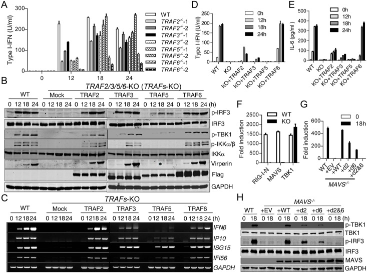
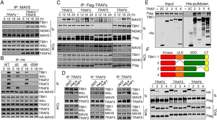
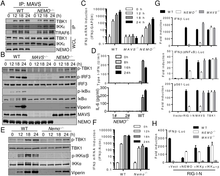
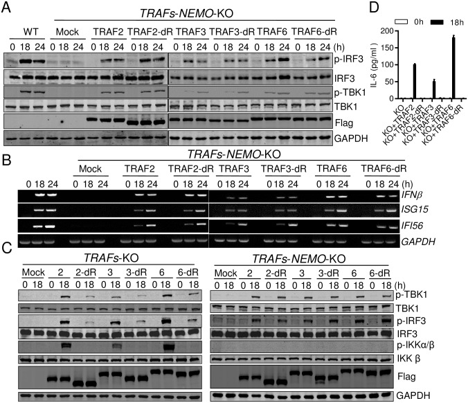
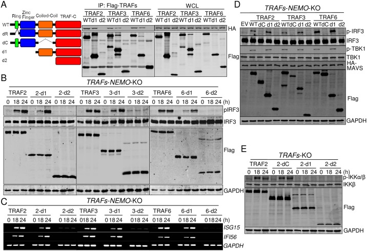
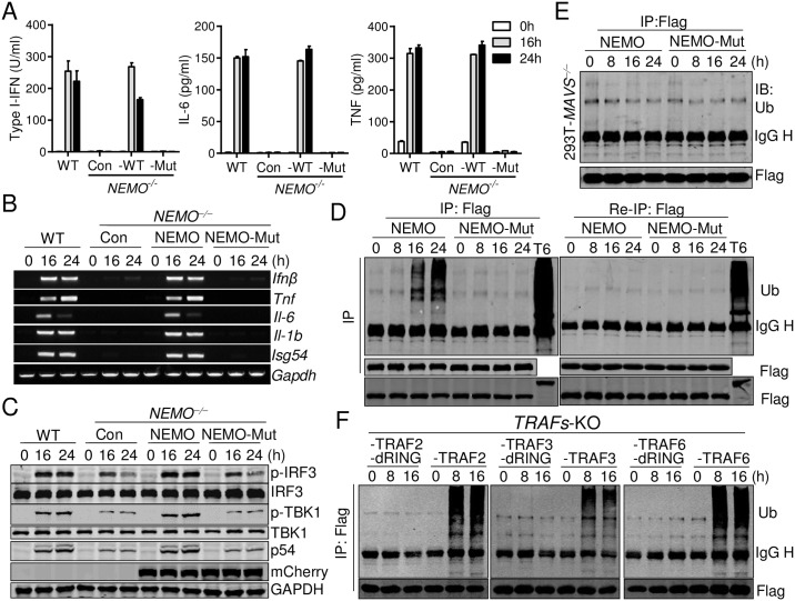
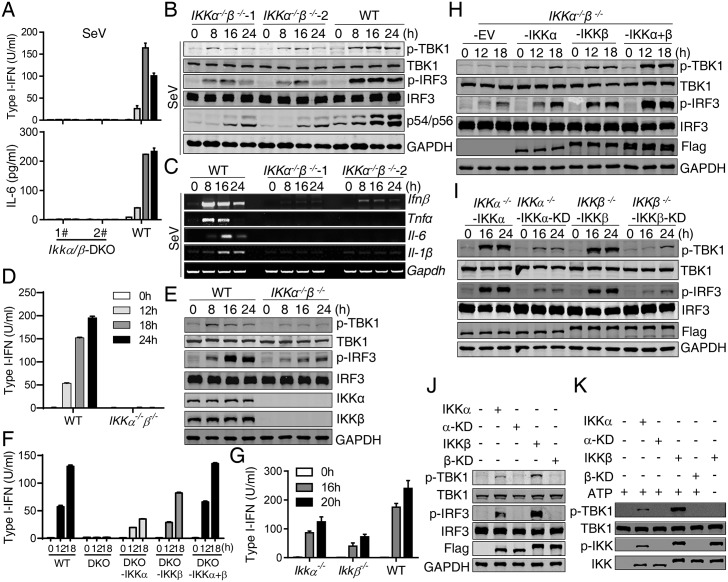
Mitochondrial antiviral-signaling protein (MAVS) transmits signals from RIG-I-like receptors after RNA virus infections. However, the mechanism by which MAVS activates downstream components, such as TBK1 and IKKα/β, is unclear, although previous work suggests the involvement of NEMO or TBK1-binding proteins TANK, NAP1, and SINTBAD. Here, we report that MAVS-mediated innate immune activation is dependent on TRAFs, partially on NEMO, but not on TBK1-binding proteins. MAVS recruited TBK1/IKKε by TRAFs that were pre-associated with TBK1/IKKε via direct interaction between the coiled-coil domain of TRAFs and the SDD domain of TBK1/IKKε. TRAF2-/-3-/-5-/-6-/- cells completely lost RNA virus responses. TRAFs' E3 ligase activity was required for NEMO activation by synthesizing ubiquitin chains that bound to NEMO for NF-κB and TBK1/IKKε activation. NEMO-activated IKKα/β were important for TBK1/IKKε activation through IKKα/β-mediated TBK1/IKKε phosphorylation. Moreover, individual TRAFs differently mediated TBK1/IKKε activation and thus fine-tuned antiviral immunity under physiological conditions.


Conflict of interest statement

The authors have declared that no competing interests exist.

Figures

Fig 1
Fig 1. TRAFs are absolutely required for MAVS-mediated signaling.
(A) WT and TRAF2−/−, TRAF3−/−, TRAF5−/−, TRAF6−/− 293T cells were infected with SeV for the indicated times. Type I-IFN production was determined by bioassay. For each knockout cell, two independent clones were accessed with similar results. (B) to (E) WT 293T cells, 293T cells deficiency of TRAF2, 3, 5, or 6 or cells deficiency of TRAF2, 3, 5, and 6 (TRAFs-deficient) reconstituted with TRAF2, 3, 5, or 6 individually were infected with SeV for the indicated times. Cells were analyzed by Western blot to detect the phosphorylation and expression of the indicated proteins (B), or RT-PCR analysis of the indicated gene induction (C), bioassay analysis of type I-IFN production (D) and ELISA analysis of IL6 production (E). (F) WT and NEMO−/− 293T cells were transfected with IFNβ–Luc reporter (50 ng) and the indicated plasmids (RIG-I-N 100 ng, MAVS 50 ng, or TBK1 50 ng). Luciferase assay was performed after 24 h. (G) to (H) WT, MAVS−/− THP1 cells and MAVS−/− THP1 cells reconstituted with MAVS-WT, MAVS-d2 (Q145N), MAVS-d6 (E155D, E457D), or MAVS-d2&6(Q145N, E155D, E457D) were infected with SeV for the indicated times. Type I-IFN production was determined by bioassay (G). Phosphorylation and expression of the indicated proteins were detected by Western blot (H). Data from (A), (D), (E), (F) and (G) represent mean ± SD. Similar results were obtained in 3 independent experiments. See also S2 Fig.
Fig 2
Fig 2. TBK1/IKKε are recruited to MAVS via the pre-associated TRAFs-TBK1/IKKe.
(A) 293T cells deficiency of TRAF2, 3, 5, or 6 or TRAFs-deficient cells reconstituted with TRAF6 were infected with SeV for the indicated times. Cell lysates were immunoprecipitated with the anti-MAVS antibody. The precipitates and whole cell lysates (WCL) were analyzed by Western blot with the indicated antibodies. (B) The reconstituted cells described in Fig 1G were infected with SeV for the indicated times. Cell lysates were immunoprecipitated with the anti-HA antibody. The precipitates and whole cell lysates (WCL) were analyzed by Western blot with the indicated antibodies. (C) The reconstituted cells described in Fig 1B were infected with SeV for the indicated times. Cell lysates were immunoprecipitated with the anti-Flag antibody. The precipitates and whole cell lysates (WCL) were analyzed by Western blot with the indicated antibodies. (D) Cell lysates of 293T, HeLa, and THP1 cells were immunoprecipitated with the anti-TRAF2, 3, or 6 antibodies. The precipitates and whole cell lysates (WCL) were analyzed by Western blot with the indicated antibodies. (E) Recombinant TBK1-Flag and His-tagged TRAFs (TRAF2C: containing amino acids 349–496) were pulled down with Ni-NTA beads. The precipitates and input were analyzed by Western blot with the indicated antibodies. (F) 293T cells were transfected with Flag-tagged TRAFs and full length TBK1 or TBK1 truncations illustrated in the upper panel for 24 h. Cell lysates were immunoprecipitated with the anti-Flag antibody. The precipitates and whole cell lysates (WCL) were analyzed by Western blot with the indicated antibodies. Truncations 1 to 4 indicate full length TBK1 and TBK1 lacking amino acids 309–385, 618–657 and 658–745. See also S3 Fig.
Fig 3
Fig 3. MAVS activates TBK1/IKKε in both NEMO-dependent and independent manner.
(A) WT and NEMO−/− 293T cells were infected with SeV for the indicated times. Cell lysates were immunoprecipitated with the anti-MAVS antibody. The precipitates and whole cell lysates (WCL) were analyzed by Western blot with the indicated antibodies. (B) and (C) WT, MAVS−/− and NEMO−/− 293T cells were infected with SeV for the indicated times. Cells were analyzed by Western blot to detect the phosphorylation and expression of the indicated proteins (B), or Quantitative–PCR analysis of Ifnβ induction (C). (D) WT and NEMO−/− HeLa cells were infected with SeV for the indicated times. Type I-IFN and IL-6 production was determined by bioassay or ELISA respectively. (E) and (F) WT and Nemo−/− MEFs were infected with SeV for the indicated times. Cells were analyzed by Western blot to detect the phosphorylation and expression of the indicated proteins (E), or Quantitative–PCR analysis of Ifnβ induction (F). (G) WT, MAVS−/− and NEMO−/− 293T cells were transfected with luciferase reporter constructs (IFNβ–Luc or IFNβ (dNF-κB)-Luc or p561-Luc, 50ng) and the indicated plasmids (empty vector 100 ng, RIG-I-N 100 ng, MAVS 50 ng and TBK1 50 ng). Luciferase assay was performed after 24 h. (H) WT, MAVS−/− and NEMO−/− 293T cells were transfected with IFNβ–Luc reporter (50 ng) and the indicated plasmids (RIG-I-N 100 ng, empty vector 200ng, NEMO 200 ng, IKKβ 200 ng, IKKα+β 100 ng each). Luciferase assay was performed after 24 h. Data from (C), (D), (F), (G) and (H) represent mean ± SD. Similar results were obtained in 3 independent experiments. See also S4 Fig.
Fig 4
Fig 4. TRAFs’ E3 ligase activity is required for TBK1/IKKε activation via NEMO.
(A) and (B) WT 293T cells, 293T cells with combined deficiency of TRAFs and NEMO (TRAFs/NEMO-deficient) and the same deficient cells reconstituted with TRAF2, 3, or 6 or their dRing deletions were infected with SeV for the indicated times. Cells were analyzed by Western blot to detect the phosphorylation and expression of the indicated proteins (A), or RT-PCR analysis of the indicated gene induction (B). (C) WT 293T cells, TRAFs-deficient and TRAFs/NEMO-deficient cells were transiently transfected with TRAF2, 3, or 6 or their dRing deletions for 12 h and infected with SeV for the indicated times. Cells were analyzed by Western blot to detect the phosphorylation and expression of the indicated proteins. (D) Cells in the left panel of (C) were analyzed by ELISA to detect the production of IL6. Data from (D) represent mean ± SD. Similar results were obtained in 3 independent experiments. See also S5 Fig.
Fig 5
Fig 5. TRAFs’ coiled-coil domain is important for both interaction and activation of TBK1/IKKε.
(A) Left: A diagram illustrating TRAF truncations. dR deletions: lacking amino acids 34–73 for TRAF2, 68–77 for TRAF3 and 70–109 for TRAF6; dC deletions: lacking amino acids 299–348 for TRAF2, 267–338 for TRAF3 and 288–348 for TRAF6; d1 deletions: lacking amino acids 1–233 for TRAF2, 1–249 for TRAF3 and 1–259 for TRAF6; d2 deletions: lacking amino acids 1–348 for TRAF2, 1–338 for TRAF3 and 1–347 for TRAF6. Right: 293T cells were transfected with Flag-tagged TRAFs or their truncations and HA-tagged TBK1 for 24 h. Cell lysates were immunoprecipitated with the anti-Flag antibody. The precipitates and whole cell lysates (WCL) were analyzed by Western blot with the indicated antibodies. (B) and (C) TRAFs/NEMO-deficient cells reconstituted with TRAF2, 3, or 6 or their truncations were infected with SeV for the indicated times. Cells were analyzed by Western blot to detect the phosphorylation and expression of the indicated proteins (B), or RT-PCR analysis of the indicated gene induction (C). (D) TRAFs/NEMO-deficient cells were transfected with Flag-tagged TRAFs or their truncations and HA-tagged MAVS for 24 h. Cells were analyzed by Western blot to detect the phosphorylation and expression of the indicated proteins. (E) TRAFs–deficient cells reconstituted with TRAF2 or its truncations were infected with SeV for the indicated times. Cells were analyzed by Western blot to detect the phosphorylation and expression of the indicated proteins.
Fig 6
Fig 6. NEMO is activated through the binding of ubiquitin chains synthetized by TRAFs.
(A-C) WT, NEMO−/− and NEMO−/− THP-1 cells reconstituted with mCherry-tagged NEMO or mCherry-tagged NEMO-Mut (Y308S/D311N/H413A/C417A) were infected with SeV for the indicated times. Type I-IFNs, IL-6 and TNF were determined by bioassay or ELISA (A). The indicated gene induction was analyzed by RT-PCR (B). Cell lysates were analyzed by Western blot to detect the phosphorylation and expression of the indicated proteins (C). (D) 293T cells stably expressing Flag-tagged NEMO or Flag-tagged NEMO-Mut were infected with SeV for the indicated times and 293T cells were transiently transfected with Flag-TRAF6 for 24 h. Cell lysates were immunoprecipitated with the anti-Flag antibody. Half of the precipitates were subjected to a second-round immunoprecipitation. The precipitates were analyzed by Western blot with the anti-ubiquitin and ant-Flag antibodies (Upper panels). The whole cell lysates (WCL) were analyzed with the anti-Flag antibody to detect the expression of Flag-tagged NEMO and TRAF6 (Bottom). (E) to (F) The same experiment was performed as in (D) except that 293T MAVS−/− cells stably expressing Flag-NEMO or Flag-NEMO-Mut (E) or TRAFs-deficient cells reconstituted with TRAF2, 3, or 6 or TRAF2/3/6-dR stably expressing Flag-tagged NEMO (F) were used. The precipitates were analyzed by Western blot with the anti-ubiquitin (Top) and ant-Flag (Bottom) antibodies. Data from (A) represent mean ± SD. Similar results were obtained in 3 independent experiments.
Fig 7
Fig 7. IKKα/β are critical for MAVS-mediated TBK1/IKKε activation.
(A) to (C) WT and IKKα−/−IKKβ−/− HeLa cells were infected with SeV for the indicated times. Type I-IFN and IL-6 production was determined by bioassay or ELISA respectively (A). Cell lysates were analyzed by Western blot to detect the phosphorylation and expression of the indicated proteins (B). The indicated gene induction was analyzed by RT-PCR (C). (D) to (E) WT and IKKα−/−IKKβ−/−THP1 cells were infected with SeV for the indicated times. Type I-IFN production was determined by bioassay (D). Cell lysates were analyzed by Western blot to detect the phosphorylation and expression of the indicated proteins (E). (F) WT, IKKα−/−IKKβ−/− and IKKα−/−IKKβ−/− HeLa cells reconstituted with IKKα, IKKβ or IKKα/β together were infected with SeV for the indicated times. Type I-IFN production was determined by bioassay. (G) WT, IKKα−/− and IKKβ−/− HeLa cells were infected with SeV for the indicated times. Type I-IFN production was determined by bioassay. (H) IKKα−/−IKKβ−/− HeLa cells reconstituted with empty vector (EV), IKKα, IKKβ or IKKα/β together were infected with SeV for the indicated times. Cell lysates were analyzed by Western blot to detect the phosphorylation and expression of the indicated proteins. (I) IKKα−/− HeLa cells reconstituted with IKKα or IKKα-KD (IKKα-D144A) and IKKβ−/− HeLa cells reconstituted with IKKβ or IKKβ-KD (IKKβ-D145A) were infected with SeV for the indicated times. Cell lysates were analyzed by Western blot to detect the phosphorylation and expression of the indicated proteins. (J) 293T cells were transfected with the indicated plasmids for 24 h. Cell lysates were analyzed by Western blot to detect the phosphorylation of the indicated proteins. (K) Flag-tagged IKKα, IKKα-KD, IKKβ or IKKβ-KD protein, stably expressed in IKKα−/−IKKβ−/− HeLa cells, was immunoprecipitated after cells were infected with SeV for 18 h, and incubated with recombinant TBK1 at room temperate for 30 min, the phosphorylation of the indicated proteins was detected by Western blot. Data from (A), (D), (F) and (G) represent mean ± SD. Similar results were obtained in 3 independent experiments. See also S7 Fig.

Similar articles

Cited by

References

    1. Takeuchi O, Akira S. Pattern Recognition Receptors and Inflammation. Cell. 2010;140(6):805–20. doi: 10.1016/j.cell.2010.01.022 - DOI - PubMed
    1. Yoneyama M, Kikuchi M, Natsukawa T, Shinobu N, Imaizumi T, Miyagishi M, et al. The RNA helicase RIG-I has an essential function in double-stranded RNA-induced innate antiviral responses. Nature immunology. 2004;5(7):730–7. Epub 2004/06/23. doi: 10.1038/ni1087 . - DOI - PubMed
    1. Gitlin L, Barchet W, Gilfillan S, Cella M, Beutler B, Flavell RA, et al. Essential role of mda-5 in type I IFN responses to polyriboinosinic:polyribocytidylic acid and encephalomyocarditis picornavirus. Proceedings of the National Academy of Sciences of the United States of America. 2006;103(22):8459–64. Epub 2006/05/23. doi: 10.1073/pnas.0603082103 . - DOI - PMC - PubMed
    1. Zust R, Cervantes-Barragan L, Habjan M, Maier R, Neuman BW, Ziebuhr J, et al. Ribose 2'-O-methylation provides a molecular signature for the distinction of self and non-self mRNA dependent on the RNA sensor Mda5. Nat Immunol. 2011;12(2):137–43. doi: 10.1038/ni.1979 - DOI - PMC - PubMed
    1. Satoh T, Kato H, Kumagai Y, Yoneyama M, Sato S, Matsushita K, et al. LGP2 is a positive regulator of RIG-I- and MDA5-mediated antiviral responses. Proc Natl Acad Sci U S A. 2010;107(4):1512–7. doi: 10.1073/pnas.0912986107 - DOI - PMC - PubMed

MeSH terms

Substances

LinkOut - more resources