

Frontal Plane Knee Mechanics and Early Cartilage Degeneration in People With Anterior Cruciate Ligament Reconstruction: A Longitudinal Study
- PMID: 29125920
- PMCID: PMC6709529
- DOI: 10.1177/0363546517739605
Frontal Plane Knee Mechanics and Early Cartilage Degeneration in People With Anterior Cruciate Ligament Reconstruction: A Longitudinal Study
Abstract
Background: Abnormal frontal plane gait mechanics are known risk factors for knee osteoarthritis, but their role in early cartilage degeneration after anterior cruciate ligament reconstruction (ACLR) is not well understood. Hypothesis/Purpose: The objective was to evaluate the association of frontal plane gait mechanics with medial knee cartilage magnetic resonance (MR) relaxation times over 1 year in patients with ACLR and controls. It was hypothesized that (1) there will be an increase in frontal plane medial knee loading and medial knee MR relaxation times over time in the patients with ACLR, and (2) increases in frontal plane medial knee loading will be associated with an increase in medial knee MR relaxation times.
Study design: Case-control study; Level of evidence, 3.
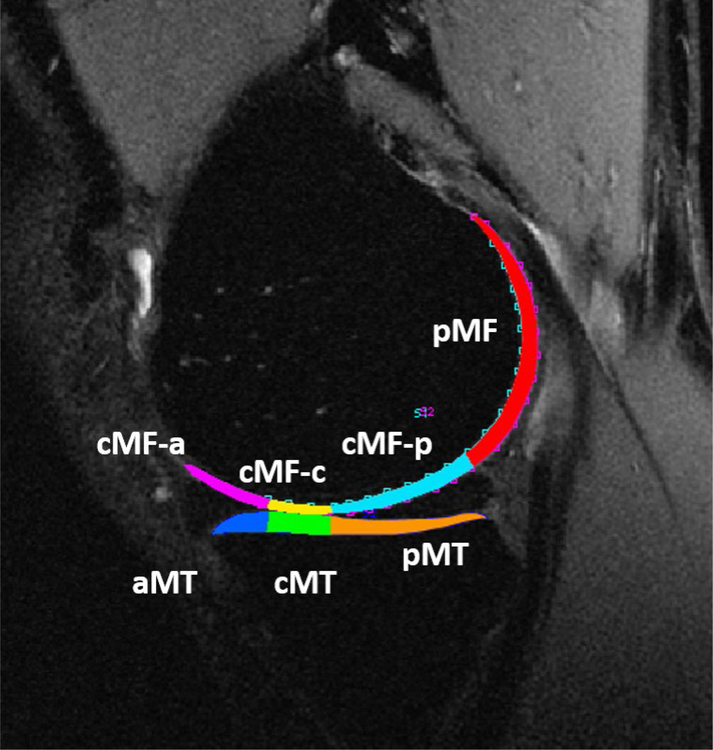
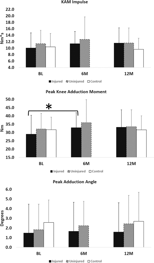
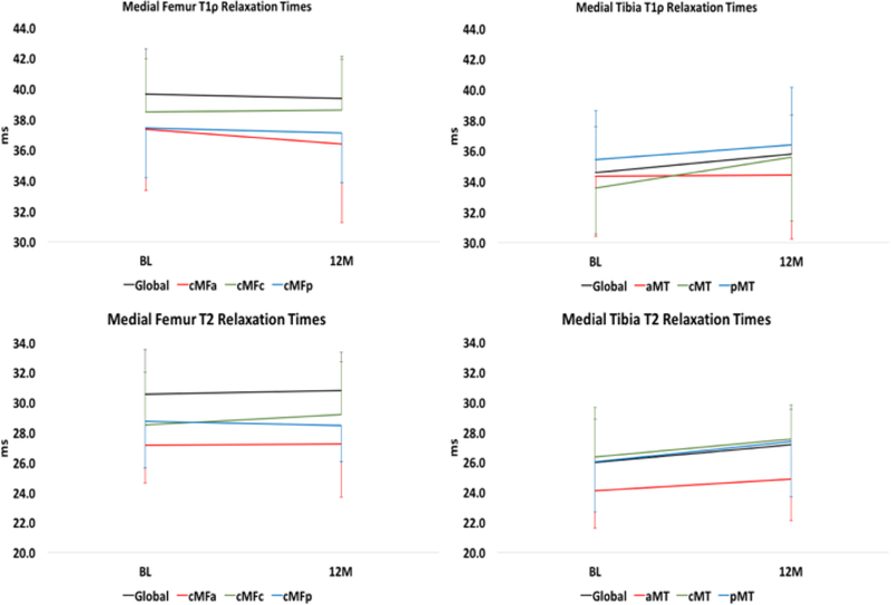
Methods: Patients with ACLR (n = 37) underwent walking gait analyses and bilateral quantitative MR imaging (MRI) before surgery and at 6 and 12 months after ACLR. Healthy control participants (n = 13) were evaluated at baseline and 12 months. Gait variables included peak knee adduction moment (KAM), KAM impulse, and peak knee adduction angle. MRI variables included medial femur and medial tibia whole compartment and subregional T1ρ and T2 relaxation times. Statistical analyses included a comparison of changes over time for gait and MRI variables, correlations between changes in gait and MRI variables over time, and differences in change in MRI variables in patients who showed an increase versus decrease in KAM impulse.
Results: There were significant increases in medial T1ρ (Δ 4%-11%) and T2 (Δ 2%-10%) relaxation times from baseline to 6 months for both knees in the ACLR group and in KAM (Δ 13%) for the injured knee. From baseline to 6 months, patients who had an increase in KAM impulse in the injured knee had a greater increase in medial T1ρ and T2 relaxation times as compared with those who did not have an increase in KAM impulse. Longitudinal changes for the control group were not significant.
Conclusion: There is an increase in medial knee relaxation times over the first 6 months after ACLR. People with an increase in medial knee loading show an increase in medial knee relaxation times when compared with those who do not have an increase in medial knee loading over the first 6 months.
Keywords: ACL; MRI; cartilage; gait; osteoarthritis.
Figures
Similar articles
-
Gait Characteristics Associated With a Greater Increase in Medial Knee Cartilage T1ρ and T2 Relaxation Times in Patients Undergoing Anterior Cruciate Ligament Reconstruction.Am J Sports Med. 2017 Dec;45(14):3262-3271. doi: 10.1177/0363546517723007. Epub 2017 Sep 12. Am J Sports Med. 2017. PMID: 28898105 Clinical Trial.
-
Cartilage Subsurface Changes to Magnetic Resonance Imaging UTE-T2* 2 Years After Anterior Cruciate Ligament Reconstruction Correlate With Walking Mechanics Associated With Knee Osteoarthritis.Am J Sports Med. 2018 Mar;46(3):565-572. doi: 10.1177/0363546517743969. Epub 2018 Jan 2. Am J Sports Med. 2018. PMID: 29293364 Free PMC article.
-
Frontal plane knee mechanics and medial cartilage MR relaxation times in individuals with ACL reconstruction: A pilot study.Knee. 2014 Oct;21(5):881-5. doi: 10.1016/j.knee.2014.06.005. Epub 2014 Jun 20. Knee. 2014. PMID: 24993277 Free PMC article.
-
Progressive Changes in Walking Kinematics and Kinetics After Anterior Cruciate Ligament Injury and Reconstruction: A Review and Meta-Analysis.J Athl Train. 2017 Sep;52(9):847-860. doi: 10.4085/1062-6050-52.6.06. J Athl Train. 2017. PMID: 28985125 Free PMC article. Review.
-
MRI T2 and T1ρ relaxation in patients at risk for knee osteoarthritis: a systematic review and meta-analysis.BMC Musculoskelet Disord. 2019 May 1;20(1):182. doi: 10.1186/s12891-019-2547-7. BMC Musculoskelet Disord. 2019. PMID: 31039785 Free PMC article.
Cited by
-
Acute Effects of Barefoot and Minimalist Footwear on Medial Tibiofemoral Compartment Loading During Running: A Statistical Parametric Mapping Approach.J Hum Kinet. 2018 Dec 31;65:35-44. doi: 10.2478/hukin-2018-0037. eCollection 2018 Dec. J Hum Kinet. 2018. PMID: 30687417 Free PMC article.
-
Effect of sensor number and location on accelerometry-based vertical ground reaction force estimation during walking.PLOS Digit Health. 2024 May 14;3(5):e0000343. doi: 10.1371/journal.pdig.0000343. eCollection 2024 May. PLOS Digit Health. 2024. PMID: 38743651 Free PMC article.
-
Acutely Normalizing Walking Speed Does Not Normalize Gait Biomechanics Post-Anterior Cruciate Ligament Reconstruction.Med Sci Sports Exerc. 2024 Mar 1;56(3):464-475. doi: 10.1249/MSS.0000000000003330. Epub 2023 Nov 27. Med Sci Sports Exerc. 2024. PMID: 38051127 Free PMC article.
-
Cartilage compositional MRI-a narrative review of technical development and clinical applications over the past three decades.Skeletal Radiol. 2024 Sep;53(9):1761-1781. doi: 10.1007/s00256-024-04734-z. Epub 2024 Jul 9. Skeletal Radiol. 2024. PMID: 38980364 Free PMC article. Review.
-
Delayed cartilage oligomeric matrix protein response to loading is associated with femoral cartilage composition post-ACLR.Eur J Appl Physiol. 2023 Nov;123(11):2525-2535. doi: 10.1007/s00421-023-05253-w. Epub 2023 Jun 16. Eur J Appl Physiol. 2023. PMID: 37326876 Free PMC article.
References
-
- Ajuied A, Wong F, Smith C, et al. Anterior cruciate ligament injury and radiologic progression of knee osteoarthritis: a systematic review and meta-analysis. Am J Sports Med 2014;42(9):2242–2252. - PubMed
-
- Akella SV, Regatte RR, Gougoutas AJ, et al. Proteoglycan-induced changes in T1rho-relaxation of articular cartilage at 4T. Magn Reson Med 2001;46(3):419–423. - PubMed
-
- Benjamini Y, Hochberg Y. Controlling the False Discovery Rate - a Practical and Powerful Approach to Multiple Testing. Journal of the Royal Statistical Society Series B-Methodological 1995;57(1):289–300.
-
- Butler RJ, Minick KI, Ferber R, Underwood F. Gait mechanics after ACL reconstruction: implications for the early onset of knee osteoarthritis. Br J Sports Med 2009;43(5):366–370. - PubMed
Publication types
MeSH terms
Grants and funding
LinkOut - more resources
Full Text Sources
Other Literature Sources
Medical
