Review
. 2018 Apr:49:16-23.
doi: 10.1016/j.conb.2017.10.020. Epub 2017 Nov 8.

Gating of visual processing by physiological need

Affiliations
Review

Gating of visual processing by physiological need

Christian R Burgess et al. Curr Opin Neurobiol. 2018 Apr.

Abstract

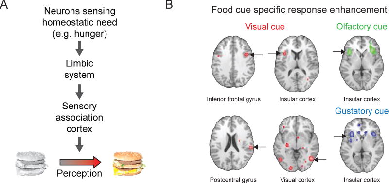
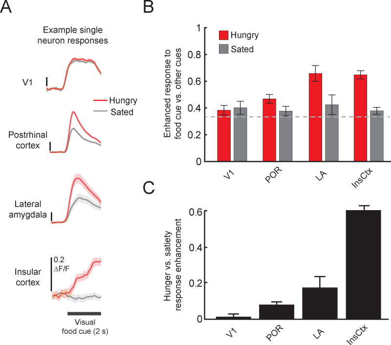
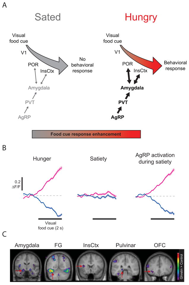
Physiological need states and associated motivational drives can bias visual processing of cues that help meet these needs. Human neuroimaging studies consistently show a hunger-dependent, selective enhancement of responses to images of food in association cortex and amygdala. More recently, cellular-resolution imaging combined with circuit mapping experiments in behaving mice have revealed underlying neuronal population dynamics and enabled tracing of pathways by which hunger circuits influence the assignment of value to visual objects in visual association cortex, insular cortex, and amygdala. These experiments begin to provide a mechanistic understanding of motivation-specific neural processing of need-relevant cues in healthy humans and in disease states such as obesity and other eating disorders.


Conflict of interest statement

Conflicts of interest: none

The authors have no conflicts of interest to disclose

Figures

Figure 1
Figure 1. Physiological needs bias processing of salient visual cues
A. To help meet the changing needs of our bodies, we direct our attention and neural processing towards relevant sensory cues in the environment. A specific physiological or homeostatic need is sensed by specialized neurons in the brain that coordinate complex search and consummatory behaviors to satisfy the need. This need state is relayed through limbic structures to cortical areas that process environmental stimuli, enhancing representations of salient objects (i.e., those relevant to the current physiological need). For example, hunger is a powerful homeostatic drive that biases visual processing towards food-associated sensory cues, such as images of cheeseburgers. B. A meta-analysis across human neuroimaging studies shows hunger-dependent enhancement of responses to visual food cues (red regions, left and middle columns) in many brain areas, including in association cortex (fusiform gyrus and parahippocampal gyrus) and insular cortex. Responses to other stimuli predicting food, including odors (top right) and tastes (bottom right), are also affected by hunger state in insular cortex. Pseudocolored pixels indicate brain regions with significant response enhancement in states of hunger vs. satiety. Modified from Obesity, 22, C. Huerta, P. Sarkar, T. Duong, A. Laird, and P. Fox, Neural bases of food perception: Coordinate-based meta-analyses of neuroimaging studies in multiple modalities, 1439–1446, 2014, with permission from John Wiley and Sons.
Figure 2
Figure 2. Cellular imaging of enhanced responses to food cues in food-restricted mice
A. Average food cue-evoked calcium response timecourses using two-photon imaging in mouse primary visual cortex (V1), postrhinal cortex (POR), lateral amygdala (LA; recorded from lateral amygdala axons in postrhinal cortex), and insular cortex (InsCtx). Neurons in POR, LA, and InsCtx tended to show increased activity in response to the same visual food-predicting cue when mice were hungry vs. after a period of feeding to satiety. ΔF/F: fractional change in fluorescence of the GCaMP6 calcium indicator. Mean +/− s.e.m. across trials. Reprinted from Neuron 91, C. Burgess, R. Ramesh, A. Sugden, K. Levandowski, M. Minnig, H. Fenselau, B. Lowell, and M. Andermann, Hunger-Dependent Enhancement of Food Cue Responses in Mouse Postrhinal Cortex and Lateral Amygdala, 1154–1169, 2016 with permission from Elsevier. B. At the population level, V1 did not show a bias towards food cues vs. neutral or aversive cues (food cue bias = (food cue response)/(sum of responses to all 3 visual cues); no bias = 0.33), regardless of hunger state. However, POR, LA, and InsCtx all showed a bias towards the food cue when the mice were hungry. Similar to findings from human neuroimaging studies, this bias was not present when mice were sated. C. Food cue response enhancement between sated and hungry states increases from V1 to POR, LA, and InsCtx. Hunger-satiety modulation index = (ResponseHungry − ResponseSated)/(ResponseHungry + ResponseSated).
Figure 3
Figure 3. Circuits regulating hunger-dependent enhancement of responses to visual food cues
A. Circuit mapping experiments in mice have begun to trace the network of brain areas responsible for hunger-dependent visual processing of food-associated cues. Agouti-related peptide (AgRP) neurons in the arcuate nucleus of the hypothalamus respond to caloric deficiency and drive feeding-related behaviors. This hunger drive is transmitted via the paraventricular thalamus (PVT) to the basolateral amygdala, which in turn exhibits reciprocal connections with postrhinal cortex (POR) and insular cortex (InsCtx). This connectivity integrates information about hunger state with the learned value of food-predicting cues, thereby enhancing responses to motivationally-salient visual stimuli and guiding behavioral choices upon presentation of food cues. Similar circuitry may be recruited by other need states (e.g. thirst, salt appetite) and may be hijacked by maladaptive drives, such as during drug seeking. B. Pharmacogenetic activation of AgRP neurons in sated mice was sufficient to restore InsCtx neuronal responses to visual food cues to levels observed in hungry mice. Subsets of InsCtx neurons responded to a visual food cue with either an increase (magenta) or decrease (blue) in activity when the mice were hungry. These responses were largely abolished when the mice were sated, but could be restored by activation of AgRP neurons. Reprinted with permission from Nature Publishing Group. C. Ghrelin administration in humans, which induces hunger, is also sufficient to induce enhanced responses to food cues vs. neutral cues, likely through actions on AgRP neurons. Colored pixels indicate the t-statistic for regions with significantly stronger responses to food images vs. scenery. Modified from Cell Metabolism, 7, S. Malik, F. McGlone, D Bedrossian and A. Dagher, Ghrelin modulates brain activity in areas that control appetitive behavior, 400–408, 2008, with permission from Elsevier. V1: primary visual cortex, FG: fusiform gyrus, OFC: orbitofrontal cortex.

References

    1. Castellanos EH, Charboneau E, Dietrich MS, Park S, Bradley BP, Mogg K, Cowan RL. Obese adults have visual attention bias for food cue images: evidence for altered reward system function. Int J Obes (Lond) 2009;33:1063–1073. - PubMed
    1. Loeber S, Grosshans M, Herpertz S, Kiefer F, Herpertz SC. Hunger modulates behavioral disinhibition and attention allocation to food-associated cues in normal-weight controls. Appetite. 2013;71:32–39. - PubMed
    1. Piech RM, Pastorino MT, Zald DH. All I saw was the cake. Hunger effects on attentional capture by visual food cues. Appetite. 2010;54:579–582. - PubMed
    1. Cornier MA. The effects of overfeeding and propensity to weight gain on the neuronal responses to visual food cues. Physiol Behav. 2009;97:525–530. - PMC - PubMed
    1. Cornier MA, Von Kaenel SS, Bessesen DH, Tregellas JR. Effects of overfeeding on the neuronal response to visual food cues. Am J Clin Nutr. 2007;86:965–971. - PubMed

Publication types

LinkOut - more resources