

Histomorphological and biochemical properties of plantar soft tissue in diabetes
- PMID: 29126035
- PMCID: PMC5937986
- DOI: 10.1016/j.foot.2017.06.001
Histomorphological and biochemical properties of plantar soft tissue in diabetes
Abstract
Background: Diabetes results in pathophysiological changes, leading to tissue that is unable to withstand and adapt to the same loads, resulting in breakdown. Certain locations are more susceptible to breakdown, yet differences between locations are largely not well understood. The authors performed a histological and biochemical analysis of isolated plantar adipose tissue at six relevant locations.
Methods: Tissue from six plantar locations (hallux, first, third and fifth metatarsal heads, lateral midfoot and calcaneus) was taken from fresh cadaveric feet of older diabetic and older non-diabetic intact donors. Histomorphological and biochemical analysis of isolated plantar tissue from both diabetic and non-diabetic feet at six relevant locations was performed.
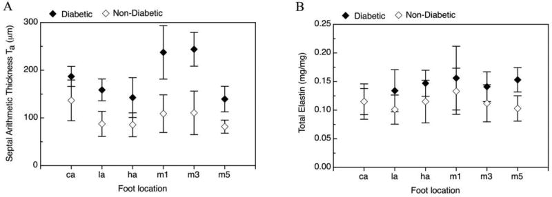
Results: The main differences found between diabetic and non-diabetic tissue were in the thickness of the septal walls and the elastin content. Diabetic tissue had significantly thicker septal walls and an increased elastin concentration. When comparing the calcaneus to other locations, although there were no differences found in the thickness of the septal walls of diabetic tissue, elastin content was lower in the calcaneous tissue compared to the non-calcaneus sites.
Conclusions: Modifications in the structural and biochemical properties could translate to changes in the mechanical properties. This information could lead to an understanding of how the structural and biochemical changes result in an increase in susceptibility of tissue to breakdown with load at the different locations of the foot.
Keywords: Biochemical; Diabetic foot ulcer; Histomorphological; Plantar soft tissue.
Copyright © 2017 Elsevier Ltd. All rights reserved.
Conflict of interest statement
The authors have no conflicts of interest to declare
Figures
Similar articles
-
The compressive mechanical properties of diabetic and non-diabetic plantar soft tissue.J Biomech. 2010 Jun 18;43(9):1754-60. doi: 10.1016/j.jbiomech.2010.02.021. Epub 2010 Mar 6. J Biomech. 2010. PMID: 20207359 Free PMC article.
-
The compressive, shear, biochemical, and histological characteristics of diabetic and non-diabetic plantar skin are minimally different.J Biomech. 2021 Dec 2;129:110797. doi: 10.1016/j.jbiomech.2021.110797. Epub 2021 Oct 7. J Biomech. 2021. PMID: 34688066 Free PMC article.
-
The shear mechanical properties of diabetic and non-diabetic plantar soft tissue.J Biomech. 2012 Jan 10;45(2):364-70. doi: 10.1016/j.jbiomech.2011.10.021. Epub 2011 Nov 12. J Biomech. 2012. PMID: 22079385 Free PMC article.
-
Can ultrasound measures of intrinsic foot muscles and plantar soft tissues predict future diabetes-related foot disease? A systematic review.PLoS One. 2018 Jun 15;13(6):e0199055. doi: 10.1371/journal.pone.0199055. eCollection 2018. PLoS One. 2018. PMID: 29906277 Free PMC article.
-
The plantar fat pad and the diabetic foot--a review.Int Wound J. 2015 Dec;12(6):636-40. doi: 10.1111/iwj.12173. Epub 2013 Oct 17. Int Wound J. 2015. PMID: 24131727 Free PMC article. Review.
Cited by
-
[Development and Validation of a Risk Prediction Model for Prolonged Hospitalization in Patients With Diabetic Foot Ulcers].Sichuan Da Xue Xue Bao Yi Xue Ban. 2024 Jul 20;55(4):972-979. doi: 10.12182/20240760507. Sichuan Da Xue Xue Bao Yi Xue Ban. 2024. PMID: 39170009 Free PMC article. Chinese.
-
Comparison of material properties of heel pad between adults with and without type 2 diabetes history: An in-vivo investigation during gait.Front Endocrinol (Lausanne). 2022 Aug 17;13:894383. doi: 10.3389/fendo.2022.894383. eCollection 2022. Front Endocrinol (Lausanne). 2022. PMID: 36060939 Free PMC article.
-
An exploratory in-situ dynamic mechanical analysis on the shearing stress-strain mechanism of human plantar soft tissue.Sci Rep. 2024 May 25;14(1):11953. doi: 10.1038/s41598-024-62713-9. Sci Rep. 2024. PMID: 38796594 Free PMC article.
-
A narrative review of the measurement methods for biomechanical properties of plantar soft tissue in patients with diabetic foot.Front Endocrinol (Lausanne). 2024 Jul 29;15:1332032. doi: 10.3389/fendo.2024.1332032. eCollection 2024. Front Endocrinol (Lausanne). 2024. PMID: 39135623 Free PMC article. Review.
-
Comparison of texture-based classification and deep learning for plantar soft tissue histology segmentation.Comput Biol Med. 2021 Jul;134:104491. doi: 10.1016/j.compbiomed.2021.104491. Epub 2021 May 15. Comput Biol Med. 2021. PMID: 34090017 Free PMC article.
References
-
- Reiber GE, Boyko EJ, Smith DG. Lower extremity foot ulcers and amputations in diabetes. In: Harris MI, editor. Diabetes in America. 2. Washington DC: U.S. Government Printing Office; 1995. pp. 409–28.
-
- Boyko EJ, Ahroni JH, Stensel V, Forsberg RC, Davignon DR, Smith DG. A prospective study of risk factors for diabetic foot ulcer. The Seattle Diabetic Foot Study. Diabetes Care. 1999;22:1036–42. - PubMed
Publication types
MeSH terms
Grants and funding
LinkOut - more resources
Full Text Sources
Other Literature Sources
Medical
