

T2*-Mapping of Acetabular Cartilage in Patients With Femoroacetabular Impingement at 3 Tesla: Comparative Analysis with Arthroscopic Findings
- PMID: 29126367
- PMCID: PMC5871124
- DOI: 10.1177/1947603517741168
T2*-Mapping of Acetabular Cartilage in Patients With Femoroacetabular Impingement at 3 Tesla: Comparative Analysis with Arthroscopic Findings
Abstract
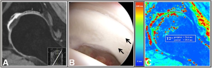
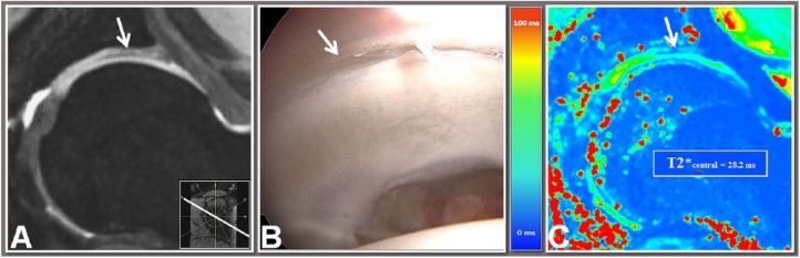
Objective To evaluate the diagnostic accuracy of T2*-mapping for detecting acetabular cartilage damage in patients with symptomatic femoroacetabular impingement (FAI). Design A total of 29 patients (17 females, 12 males, mean age 35.6 ± 12.8 years, mean body mass index 25.1 ± 4.1 kg/m2, 16 right hips) with symptomatic FAI underwent T2* MRI and subsequent hip arthroscopy. T2* values were obtained by region of interest analysis in seven radially reformatted planes around the femoral neck (anterior, anterior-superior, superior-anterior, superior, superior-posterior, posterior-superior, posterior). Intraoperatively, a modified Outerbridge classification was used for assessment of the cartilage status in each region. T2* values and intraoperative data were compared, and sensitivity, specificity, negative predictive values (NPV) and positive predictive values (PPV) as well as the correlation between T2*-mapping and intraoperative findings, were determined. The mean time interval between MRI and arthroscopy was 65.7 ± 48.0 days. Results Significantly higher T2* values were noted in arthroscopically normal evaluated cartilage than in regions with cartilage degeneration (mean T2* 25.6 ± 4.7 ms vs. 19.9 ± 4.5 ms; P < 0.001). With the intraoperative findings as a reference, sensitivity, specificity, NPV and PPV were 83.5%, 67.7%, 78.4% and 74.4%, respectively. The correlation between T2*-mapping and intraoperative cartilage status was moderate (ρ = -0.557; P < 0.001). Conclusions T2*-mapping enabled analysis of acetabular cartilage with appropriate correlation with intraoperative findings and promising results for sensitivity, specificity, PPV, and NPV in this cohort. Our results emphasize the value of T2*-mapping for the diagnosis of hip joint cartilage pathologies in symptomatic FAI.
Keywords: FAI; MRI; T2*-mapping; arthroscopy; hip.
Conflict of interest statement
Figures

References
-
- Leunig M, Ganz R. Femoroacetabular impingement. A common cause of hip complaints leading to arthrosis [in German]. Unfallchirurg. 2005;108:9-10, 12-7. - PubMed
-
- Ho CP, Ommen ND, Bhatia S, Saroki AJ, Goljan P, Briggs KK, et al. Predictive value of 3-T magnetic resonance imaging in diagnosing grade 3 and 4 chondral lesions in the hip. Arthroscopy. 2016;32:1808-13. - PubMed
-
- Zilkens C, Miese F, Kim YJ, Jager M, Mamisch TC, Hosalkar H, et al. Direct comparison of intra-articular versus intravenous delayed gadolinium-enhanced MRI of hip joint cartilage. J Magn Reson Imaging. 2014;39:94-102. - PubMed
Publication types
MeSH terms
LinkOut - more resources
Full Text Sources
Other Literature Sources
Medical
