

The adverse vascular effects of multi-walled carbon nanotubes (MWCNTs) to human vein endothelial cells (HUVECs) in vitro: role of length of MWCNTs
- PMID: 29126419
- PMCID: PMC5681822
- DOI: 10.1186/s12951-017-0318-x
The adverse vascular effects of multi-walled carbon nanotubes (MWCNTs) to human vein endothelial cells (HUVECs) in vitro: role of length of MWCNTs
Abstract
Background: Increasing evidences indicate that exposure to multi-walled carbon nanotubes (MWCNTs) could induce adverse vascular effects, but the role of length of MWCNTs in determining the toxic effects is less studied. This study investigated the adverse effects of two well-characterized MWCNTs to human umbilical vein endothelial cells (HUVECs).
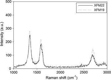
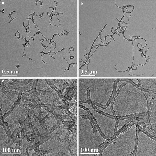
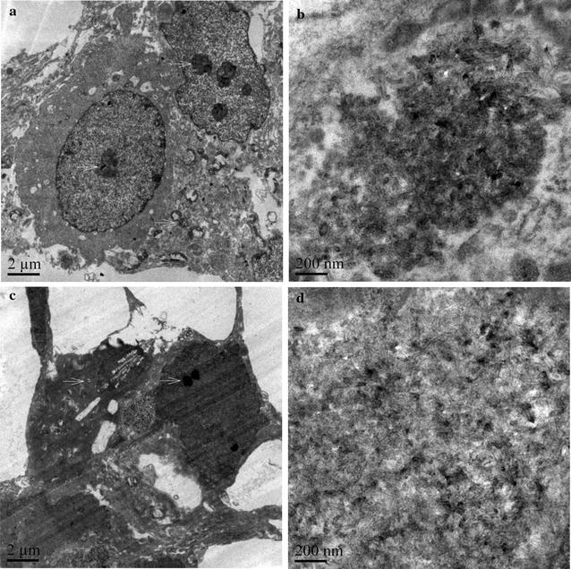
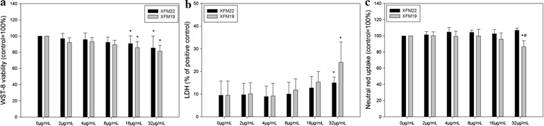
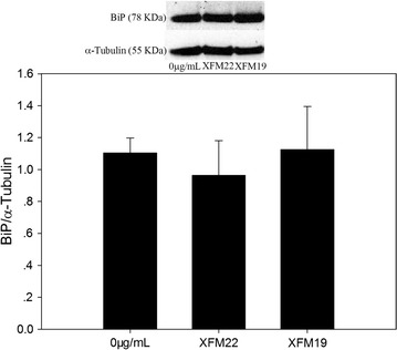
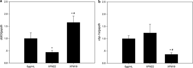
Methods: The internalization and localization of MWCNTs in HUVECs were examined by using transmission electron microscopy (TEM). The cytotoxicity of MWCNTs to HUVECs was assessed by water soluble tetrazolium-8 (WST-8), lactate dehydrogenase (LDH) and neutral red uptake assays. Oxidative stress was indicated by the measurement of intracellular glutathione (GSH) and reactive oxygen species (ROS). ELISA was used to determine the release of inflammatory cytokines. THP-1 monocyte adhesion to HUVECs was also measured. To indicate the activation of endoplasmic reticulum (ER) stress, the expression of ddit3 and xbp-1s was measured by RT-PCR, and BiP protein level was measured by Western blot.
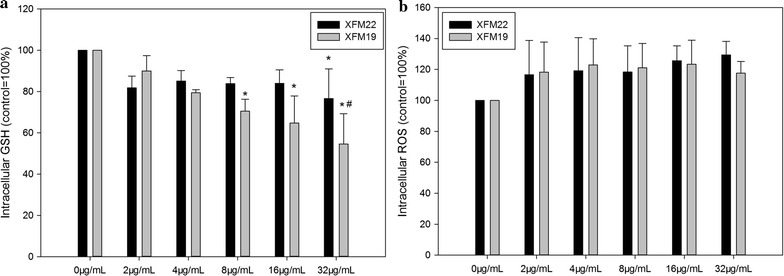
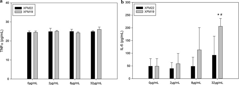
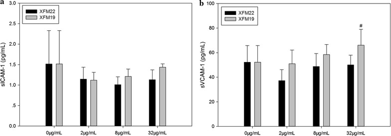
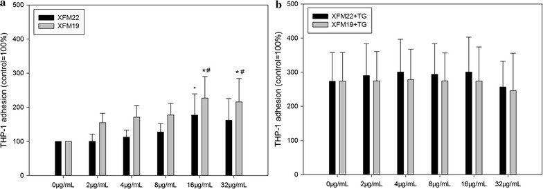
Results: Transmission electron microscopy observation indicates the internalization of MWCNTs into HUVECs, with a localization in nuclei and mitochondria. The longer MWCNTs induced a higher level of cytotoxicity to HUVECs compared with the shorter ones. Neither of MWCNTs significantly promoted intracellular ROS, but the longer MWCNTs caused a higher depletion of GSH. Exposure to both types of MWCNTs significantly promoted THP-1 adhesion to HUVECs, accompanying with a significant increase of release of interleukin-6 (IL-6) but not tumor necrosis factor α (TNFα), soluble ICAM-1 (sICAM-1) or soluble VCAM-1 (sVCAM-1). Moreover, THP-1 adhesion and release of IL-6 and sVCAM-1 induced by the longer MWCNTs were significantly higher compared with the responses induced by the shorter ones. The biomarker of ER stress, ddit3 expression, but not xbp-1s expression or BiP protein level, was significantly induced by the exposure of longer MWCNTs.
Conclusions: Combined, these results indicated length dependent toxic effects of MWCNTs to HUVECs in vitro, which might be associated with oxidative stress and activation of ER stress.
Keywords: Endoplasmic reticulum (ER) stress; Human umbilical vein endothelial cell (HUVEC); Multi-walled carbon nanobute (MWCNT); Oxidative stress; Vascular effect.
Figures
Similar articles
-
Cytotoxicity, cytokine release and ER stress-autophagy gene expression in endothelial cells and alveolar-endothelial co-culture exposed to pristine and carboxylated multi-walled carbon nanotubes.Ecotoxicol Environ Saf. 2018 Oct;161:569-577. doi: 10.1016/j.ecoenv.2018.06.025. Epub 2018 Jun 18. Ecotoxicol Environ Saf. 2018. PMID: 29929133
-
The toxicity of multi-walled carbon nanotubes (MWCNTs) to human endothelial cells: The influence of diameters of MWCNTs.Food Chem Toxicol. 2019 Apr;126:169-177. doi: 10.1016/j.fct.2019.02.026. Epub 2019 Feb 22. Food Chem Toxicol. 2019. PMID: 30802478
-
Multi-Walled Carbon Nanotubes (MWCNTs) Activate Apoptotic Pathway Through ER Stress: Does Surface Chemistry Matter?Int J Nanomedicine. 2019 Nov 28;14:9285-9294. doi: 10.2147/IJN.S217977. eCollection 2019. Int J Nanomedicine. 2019. PMID: 31819430 Free PMC article.
-
The impact of multi-walled carbon nanotubes (MWCNTs) on macrophages: contribution of MWCNT characteristics.Sci China Life Sci. 2018 Nov;61(11):1333-1351. doi: 10.1007/s11427-017-9242-3. Epub 2018 May 22. Sci China Life Sci. 2018. PMID: 29797182 Review.
-
Necrotic, apoptotic and autophagic cell fates triggered by nanoparticles.Autophagy. 2019 Jan;15(1):4-33. doi: 10.1080/15548627.2018.1509171. Epub 2018 Sep 13. Autophagy. 2019. PMID: 30160607 Free PMC article. Review.
Cited by
-
G-Optrode Bio-Interfaces for Non-Invasive Optical Cell Stimulation: Design and Evaluation.Biosensors (Basel). 2022 Sep 30;12(10):808. doi: 10.3390/bios12100808. Biosensors (Basel). 2022. PMID: 36290945 Free PMC article.
-
Toxicity of combined exposure of ZnO nanoparticles (NPs) and myricetin to Caco-2 cells: changes of NP colloidal aspects, NP internalization and the apoptosis-endoplasmic reticulum stress pathway.Toxicol Res (Camb). 2019 Jun 20;8(5):613-620. doi: 10.1039/c9tx00127a. eCollection 2019 Sep 1. Toxicol Res (Camb). 2019. PMID: 31588339 Free PMC article.
-
In vitro toxicity of carbon nanotubes: a systematic review.RSC Adv. 2022 May 31;12(25):16235-16256. doi: 10.1039/d2ra02519a. eCollection 2022 May 23. RSC Adv. 2022. PMID: 35733671 Free PMC article. Review.
-
Physicochemical characterization and genotoxicity of the broad class of carbon nanotubes and nanofibers used or produced in U.S. facilities.Part Fibre Toxicol. 2020 Dec 7;17(1):62. doi: 10.1186/s12989-020-00392-w. Part Fibre Toxicol. 2020. PMID: 33287860 Free PMC article.
-
Graphene Nanomaterials: Synthesis, Biocompatibility, and Cytotoxicity.Int J Mol Sci. 2018 Nov 12;19(11):3564. doi: 10.3390/ijms19113564. Int J Mol Sci. 2018. PMID: 30424535 Free PMC article. Review.
References
MeSH terms
Substances
Grants and funding
LinkOut - more resources
Full Text Sources
Other Literature Sources
Research Materials
Miscellaneous
