

TGFβ3 recruits endogenous mesenchymal stem cells to initiate bone regeneration
- PMID: 29126441
- PMCID: PMC5681754
- DOI: 10.1186/s13287-017-0693-0
TGFβ3 recruits endogenous mesenchymal stem cells to initiate bone regeneration
Retraction in
-
Retraction Note: TGFβ3 recruits endogenous mesenchymal stem cells to initiate bone regeneration.Stem Cell Res Ther. 2022 Aug 5;13(1):402. doi: 10.1186/s13287-022-03108-3. Stem Cell Res Ther. 2022. PMID: 35932081 Free PMC article. No abstract available.
Abstract
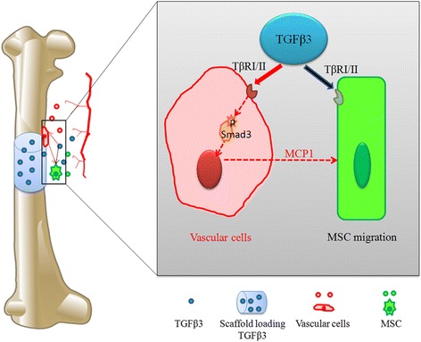
Background: The recruitment of a sufficient number of endogenous mesenchymal stem cells (MSCs) is the first stage of in-situ tissue regeneration. Transforming growth factor beta-3 (TGFβ3) could recruit stem or progenitor cells and endothelial cells to participate in tissue regeneration. However, the mechanism of TGFβ3 recruiting MSCs toward bone regeneration has remained obscure.
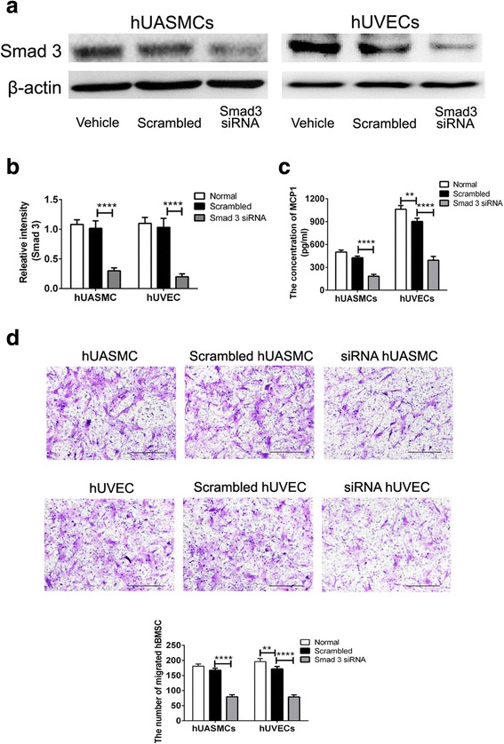
Methods: We estimated the promigratory property of TGFβ3 on human bone marrow MSCs (hBMSCs) cocultured with the vascular cells (human umbilical artery smooth muscle cells or human umbilical vein endothelial cells) or not by Transwell assay. After the addition of the inhibitor (SB431542) or Smad3 siRNA, the levels of MCP1 and SDF1 in coculture medium were tested by ELISA kit, and then the migratory signaling pathway of hBMSCs induced by TGFβ3 was investigated by western blot analysis. In vivo, a 2-mm FVB/N mouse femur defect model was used to evaluate chemokine secretion, endogenous cell homing, and bone regeneration induced by scaffolds loading 1 μg TGFβ3 through qPCR, immunofluorescent staining, immunohistochemical analysis, and Micro-CT, compared to the vehicle group.
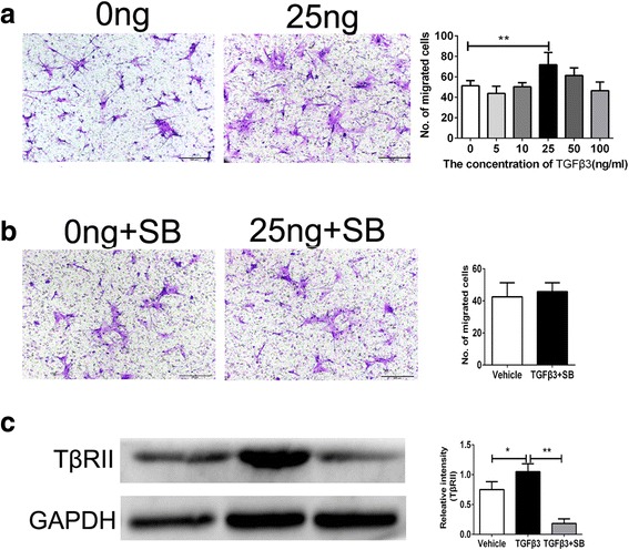
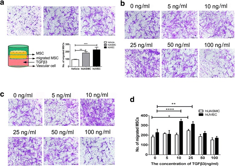
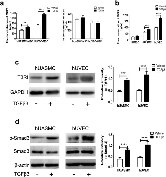
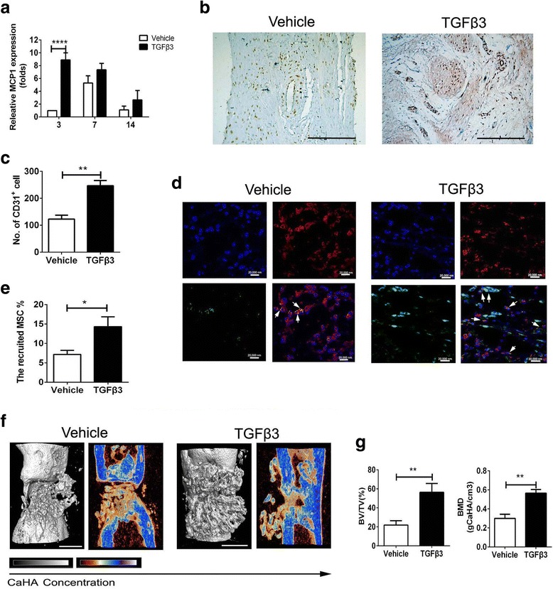
Results: TGFβ3 (25 ng/ml) directly showed a nearly 40% increase in migrated hBMSCs via the TGFβ signaling pathway, compared to the vehicle treatment. Then, in the coculture system of hBMSCs and vascular cells, TGFβ3 further upregulated nearly 3-fold MCP1 secretion from vascular cells in a Smad3-dependent manner, to indirectly enhance nearly more than 50% of migrated hBMSCs. In vivo, TGFβ3 delivery improved MCP1 expression by nearly 7.9-fold, recruited approximately 2.0-fold CD31+ vascular cells and 2.0-fold Sca-1+ PDGFR-α+ MSCs, and achieved 2.5-fold bone volume fraction (BV/TV) and 2.0-fold bone mineral density, relative to TGFβ3-free delivery.
Conclusions: TGFβ3, as a MSC homing molecule, recruited MSCs to initiate bone formation in the direct-dependent and indirect-dependent mechanisms. This may shed light on the improvement of MSC homing in bone regeneration.
Keywords: MCP1; Mesenchymal stem cell; Recruitment; TGFβ3; Vascular cells.
Conflict of interest statement
Ethics approval and consent to participate
The animal study protocol complied with the Animal Management Rule of the Ministry of Public Health, China (documentation 55, 2001).
Consent for publication
Not applicable.
Competing interests
The authors declare that they have no competing interests.
Publisher’s Note
Springer Nature remains neutral with regard to jurisdictional claims in published maps and institutional affiliations.
Figures
References
-
- Cipitria A, Boettcher K, Schoenhals S, Garske DS, Schmidt-Bleek K, Ellinghaus A, Dienelt A, Peters A, Mehta M, Madl CM, Huebsch N, Mooney DJ, Duda GN. In-situ tissue regeneration through SDF-1alpha driven cell recruitment and stiffness-mediated bone regeneration in a critical-sized segmental femoral defect. Acta Biomater. 2017;60:50. doi: 10.1016/j.actbio.2017.07.032. - DOI - PubMed
-
- Ponte AL, Marais E, Gallay N, Langonné A, Delorme B, Hérault O, Charbord P, Domenech J. The in vitro migration capacity of human bone marrow mesenchymal stem cells: comparison of chemokine and growth factor chemotactic activities. Stem Cells. 2007;25:1737. doi: 10.1634/stemcells.2007-0054. - DOI - PubMed
Publication types
MeSH terms
Substances
Grants and funding
- 31400827/National Natural Science Foundation of China
- 81472059/National Natural Science Foundation of China
- 2015AA020315/National high technology research and development program of China
- SWH2016JCYB-11/the Foundation of Southwest Hospital
- SWH2016ZDCX1015/Major Program of Southwest Hospital Foundation
LinkOut - more resources
Full Text Sources
Other Literature Sources
Research Materials
Miscellaneous
