. 2017 Nov 10;10(1):54.
doi: 10.1186/s13072-017-0160-2.

Ubiquitin C-terminal hydrolase isozyme L1 is associated with shelterin complex at interstitial telomeric sites

Affiliations

Ubiquitin C-terminal hydrolase isozyme L1 is associated with shelterin complex at interstitial telomeric sites

Aleksandar Ilic et al. Epigenetics Chromatin. .

Abstract

Background: Ubiquitin C-terminal hydrolase isozyme L1 (UCHL1) is primarily expressed in neuronal cells and neuroendocrine cells and has been associated with various diseases, including many cancers. It is a multifunctional protein involved in deubiquitination, ubiquitination and ubiquitin homeostasis, but its specific roles are disputed and still generally undetermined.

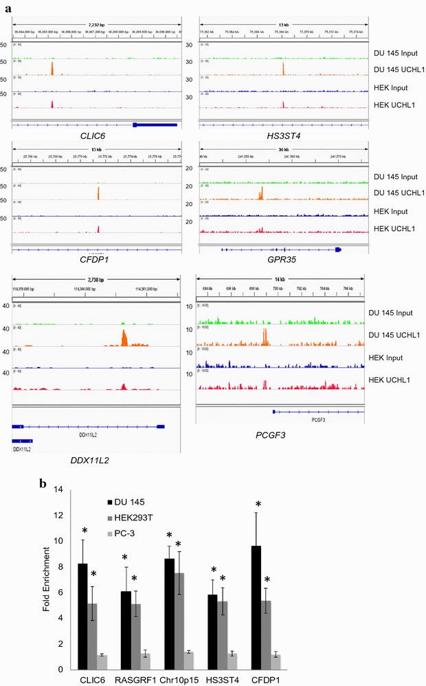
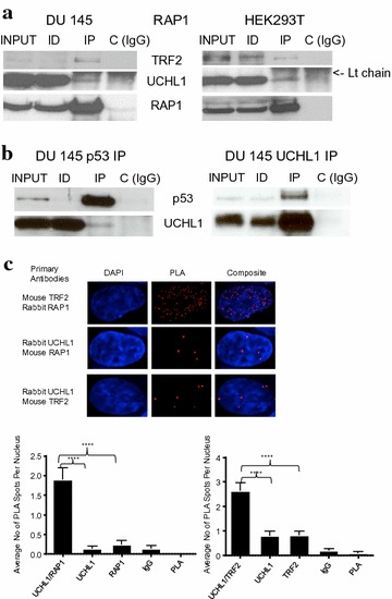
Results: Herein, we demonstrate that UCHL1 is associated with genomic DNA in certain prostate cancer cell lines, including DU 145 cells derived from a brain metastatic site, and in HEK293T embryonic kidney cells with a neuronal lineage. Chromatin immunoprecipitation and sequencing revealed that UCHL1 localizes to TTAGGG repeats at telomeres and interstitial telomeric sequences, as do TRF1 and TRF2, components of the shelterin complex. A weak or transient interaction between UCHL1 and the shelterin complex was confirmed by immunoprecipitation and proximity ligation assays. UCHL1 and RAP1, also known as TERF2IP and a component of the shelterin complex, were bound to the nuclear scaffold.

Conclusions: We demonstrated a novel feature of UCHL1 in binding telomeres and interstitial telomeric sites.

Keywords: Interstitial telomeric sites; Prostate cancer; Shelterin complex; UCHL1.


Figures

Fig. 1
Fig. 1
UCHL1 is associated with genomic DNA in prostate cancer cells expressing it. a DNA cross-linked proteins from BPH-1, DU 145, PC-3 and LNCaP cells treated with 1 mM cisplatin were electrophoretically resolved on two-dimensional PAGE. The gels were stained with silver. be Total cellular proteins (TCP) or DNA cross-linked proteins isolated by hydroxyapatite column chromatography from cells treated with 1 mM cisplatin or 1% formaldehyde were resolved by SDS-10% PAGE and immunoblotted with indicated antibodies. Cells in bd were BPH-1 (B), DU 145 (D), PC-3 (P) and LNCaP (L)
Fig. 2
Fig. 2
Genomic distribution of UCHL1 in DU 145 cells. a Chromosome distribution of UCHL1 binding sites: Blue bars represent the relative lengths of each chromosome, and red bars represent the percentage of UCHL1 peaks (out of 191 peaks) per chromosome. P value indicates the significance of the enrichment of ChIPped regions relative to the genome background and is shown in parentheses. b UCHL1 binding sites were divided according to their relative locations to gene loci
Fig. 3
Fig. 3
Representation and validation of random UCHL1 binding sites in DU 145 and HEK293T cells. a Signal tracks showing DNA enrichment in UCHL1 ChIP compared to genomic input. The peak signals were normalized by igvtools. b ChIP experiments were performed using anti-UCHL1 antibodies on DSP/formaldehyde cross-linked 300–400-bp chromatin fragments obtained by sonication from DU 145, HEK293T or PC-3 cells. Fold enrichment values were normalized to IgG ChIP values. Asterisk (*) indicate statistical significance p < 0.05
Fig. 4
Fig. 4
UCHL1 localizes to telomeric repeat sequences as do TRF1 and TRF2. a The top five motifs were identified using the MEME software. The E value is the expected number of hits by chance (the lower the E value the more significant is the motif). b Signal tracks showing DNA enrichment in UCHL1 ChIP from DU 145 and HEK293T cells and TRF1 or TRF2 ChIP from BJHELTRasmc cells (Simonet et al. [21]—21423270) were aligned for CLIC6 and FAM157A genes. Cells were treated with formaldehyde (1-DU 145 Input and UCHL1, HEK Input and UCHL1) or DSP/formaldehyde (2-DU 145 Input and UCHL1)
Fig. 5
Fig. 5
UCHL1 interacts with the shelterin complex and the nuclear scaffold. a DU 145 or HEK293T cellular lysates in a low stringency buffer were incubated with anti-TRF2 antibodies or control IgG. The immunoprecipitate (IP), and equal volumes of lysate (Input) and immunodepleted (ID) fractions were analyzed by immunoblotting with the indicated antibodies. b In left panel, DU 145 cellular lysate in a low stringency buffer was incubated with anti-p53 antibodies or control IgG. In right panel, cell lysate in a high stringency buffer of DU 145 cells treated with the cross-linker DSP was incubated with anti-UCHL1 antibodies or control IgG. The immunoblot analysis was done as in (a). c In situ PLA images of the interaction between TRF2 and RAP1 (positive control), UCHL1 and RAP1 or UCHL1 and TRF2 in HEK293T cells. PLA signals appear as discrete red dots and nuclei are visualized by DAPI (blue). A total of 30 nuclei per group were quantified. The average number of PLA foci per nucleus was graphed with error bars representing standard errors of the means. Single primary antibodies, isotype control and PLA probes only were used as negative controls as indicated. ****p < 0.0001 (determined by one-way ANOVA)
Fig. 6
Fig. 6
UCHL1 interacts with TRF2 and co-localizes with telomeres. a Double immunofluorescence displayed co-localization of TRF2 and UCHL1 in HEK293T cells. Blue—nucleus; red—UCHL1; green—TRF2; yellow—TRF2–UCHL1 co-localizing spots. b Telomeric localization of UCHL1 was determined in HEK293T cells by immuno-FISH with antibodies against UCHL1 and fluorochrome-coupled (Cy3) telomere PNA probes. c Telomeric localization of TRF2 was determined in HEK293T cells by immuno-FISH for antibodies against TRF2 and fluorochrome-coupled (Cy3) telomere PNA probes. Blue—nucleus; red—telomeres; green—UCHL1/TRF2; yellow—telomere-UCHL1/TRF2 co-localizing spots. d Thirty HEK cell nuclei per experiment were quantified. Average number of colocalized spots per nucleus was counted and graphed. Image J software was used for quantification. Error bar represents the standard error of mean
Fig. 7
Fig. 7
UCHL1 and RAP1 are associated with the nuclear scaffold. a Proteins (5 µg) from each fraction [total cellular protein (TC), total nuclear protein (TN), ammonium sulfate fraction (AS) and nuclear scaffold protein (NS)] were resolved on a SDS 15% polyacrylamide gel and stained with Coomassie Blue. The DU 145 cells were treated or not with protein–protein cross-linker DSP. b Immunoblot analysis using the indicated antibodies on TC, TN, AS and NS proteins from DU 145 cells treated or not with protein–protein cross-linker DSP. PC-3 cells were not treated with DSP. c Immunoblot analysis of UCHL1 and RAP1 in nuclear scaffold protein complexes from DSP-treated DU 145 cells. The nuclear scaffold was solubilized in SDS sample buffer in both reducing (R) and non-reducing (NR) conditions and analyzed by SDS-PAGE on a 10% gels and immunoblotting
Fig. 8
Fig. 8
UCHL1 knockdown and shelterin protein levels. HEK293T cells were transfected with GAPDH siRNA (50 nM), scrambled siRNA control (50 nM) and two UCHL1 siRNA with the final concentrations of 25 and 50 nM. Whole protein lysates were extracted from control and siRNA-transfected HEK293T cells 72 h posttransfection, and immunoblot analysis was performed with antibodies against UCHL1, RAP1, TRF2 and GAPDH. Beta actin was used as an internal control

Similar articles

Cited by

References

    1. Day IN, Thompson RJ. UCHL1 (PGP 9.5): neuronal biomarker and ubiquitin system protein. Prog Neurobiol. 2010;90:327–362. doi: 10.1016/j.pneurobio.2009.10.020. - DOI - PubMed
    1. Pfoh R, Lacdao IK, Saridakis V. Deubiquitinases and the new therapeutic opportunities offered to cancer. Endocr Relat Cancer. 2015;22:T35–T54. doi: 10.1530/ERC-14-0516. - DOI - PMC - PubMed
    1. Wilkinson KD, Lee KM, Deshpande S, Duerksen-Hughes P, Boss JM, Pohl J. The neuron-specific protein PGP 9.5 is a ubiquitin carboxyl-terminal hydrolase. Science. 1989;246:670–673. doi: 10.1126/science.2530630. - DOI - PubMed
    1. Larsen CN, Krantz BA, Wilkinson KD. Substrate specificity of deubiquitinating enzymes: ubiquitin C-terminal hydrolases. Biochemistry. 1998;37:3358–3368. doi: 10.1021/bi972274d. - DOI - PubMed
    1. Das C, Hoang QQ, Kreinbring CA, Luchansky SJ, Meray RK, Ray SS, et al. Structural basis for conformational plasticity of the Parkinson’s disease-associated ubiquitin hydrolase UCH-L1. Proc Natl Acad Sci USA. 2006;103:4675–4680. doi: 10.1073/pnas.0510403103. - DOI - PMC - PubMed

MeSH terms

Substances

Grants and funding