

Decreased neurite density within frontostriatal networks is associated with executive dysfunction in temporal lobe epilepsy
- PMID: 29126704
- PMCID: PMC5756677
- DOI: 10.1016/j.yebeh.2017.09.012
Decreased neurite density within frontostriatal networks is associated with executive dysfunction in temporal lobe epilepsy
Abstract
Objective: Executive dysfunction is observed in a sizable number of patients with refractory temporal lobe epilepsy (TLE). The frontostriatal network has been proposed to play a significant role in executive functioning, however, because of the complex architecture of these tracts, it is difficult to generate measures of fiber tract microstructure using standard diffusion tensor imaging. To examine the association between frontostriatal network compromise and executive dysfunction in TLE, we applied an advanced, multishell diffusion model, restriction spectrum imaging (RSI), that isolates measures of intraaxonal diffusion and may provide better estimates of fiber tract compromise in TLE.
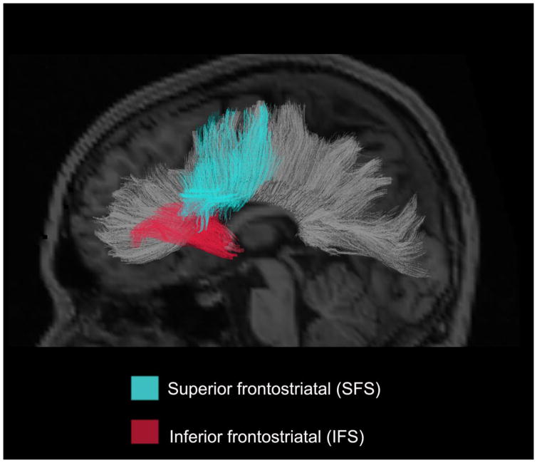
Methods: Restriction spectrum imaging scans were obtained from 32 patients with TLE [16 right TLE (RTLE); 16 left TLE (LTLE)] and 24 healthy controls (HC). An RSI-derived measure of intraaxonal anisotropic diffusion (neurite density; ND) was calculated for the inferior frontostriatal tract (IFS) and superior frontostriatal tract (SFS) and compared between patients with TLE and HC. Spearman correlations were performed to evaluate the relationships between ND of each tract and verbal (i.e., D-KEFS Category Switching Accuracy and Color-Word Interference Inhibition/Switching) and visuomotor (Trail Making Test) set-shifting performances in patients with TLE.
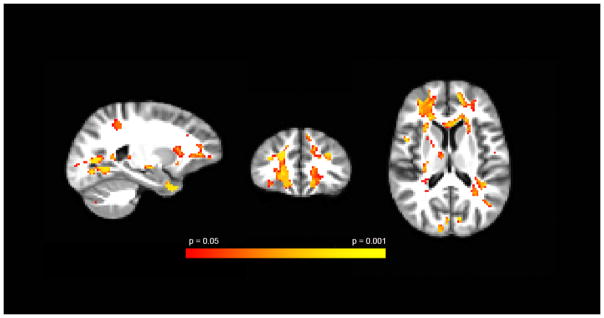
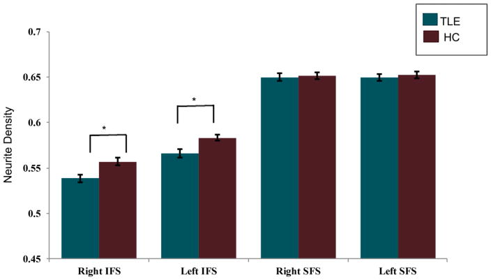
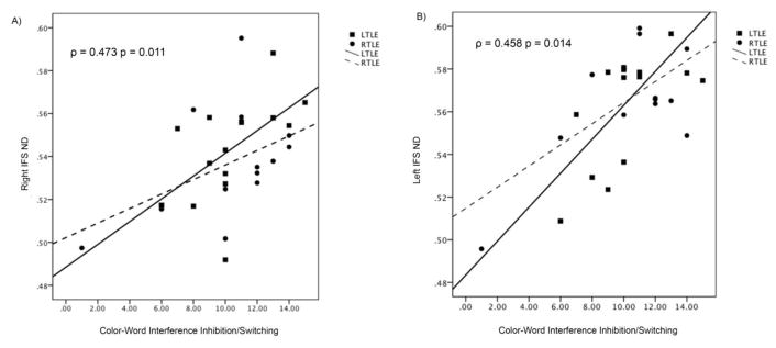
Results: Patients with TLE demonstrated reductions in ND of the left and right IFS, but not SFS, compared with HC. Reduction in ND of left and right IFS was associated with poorer performance on verbal set-shifting in TLE. Increases in extracellular diffusion (isotropic hindered; IH) were not associated with executive dysfunction in the patient group.
Significance: Restriction spectrum imaging-derived ND revealed microstructural changes within the IFS in patients with TLE, which was associated with poorer executive functioning. This suggests that axonal/myelin loss to fiber networks connecting the striatum to the inferior frontal cortex is likely contributing to executive dysfunction in TLE.
Keywords: Advanced diffusion; Demyelination; Executive function; Inhibition; Set-shifting.
Published by Elsevier Inc.
Conflict of interest statement
We confirm that we have read the Journal’s position on issues involved in ethical publication and affirm that this report is consistent with those guidelines. None of the authors have any conflicts of interest to disclose.
Figures
References
-
- Sherman EM, Slick DJ, Eyrl KL. Executive dysfunction is a significant predictor of poor quality of life in children with epilepsy. Epilepsia. 2006;47(11):1936–42. - PubMed
-
- McDonald CR, Hagler DJ, Jr, Ahmadi ME, Tecoma E, Iragui V, Gharapetian L, et al. Regional neocortical thinning in mesial temporal lobe epilepsy. Epilepsia. 2008;49(5):794–803. - PubMed
-
- Reyes A, Thesen T, Wang X, Hahn D, Yoo D, Kuzniecky R, et al. Resting-state functional MRI distinguishes temporal lobe epilepsy subtypes. Epilepsia. 2016;57(9):1475–84. - PubMed
Publication types
MeSH terms
Grants and funding
LinkOut - more resources
Full Text Sources
Other Literature Sources
Medical
