Review
. 2017 Dec:72:10-18.
doi: 10.1016/j.semcdb.2017.11.010. Epub 2017 Nov 15.

The myogenic regulatory factors, determinants of muscle development, cell identity and regeneration

Affiliations
Review

The myogenic regulatory factors, determinants of muscle development, cell identity and regeneration

J Manuel Hernández-Hernández et al. Semin Cell Dev Biol. 2017 Dec.

Abstract

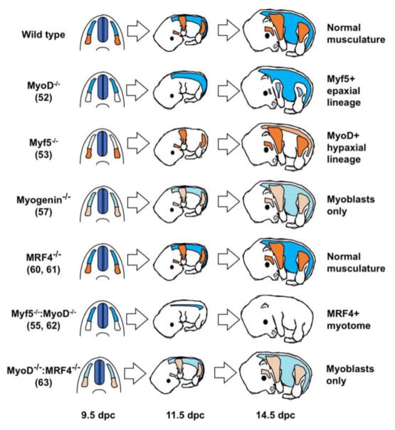
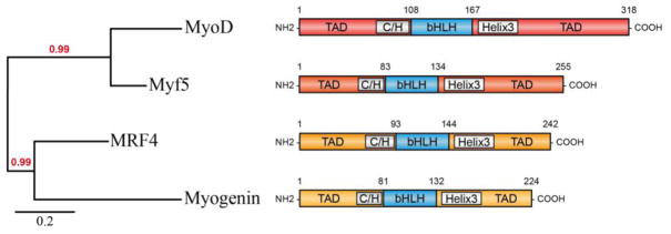
The Myogenic Regulatory Factors (MRFs) Myf5, MyoD, myogenin and MRF4 are members of the basic helix-loop-helix family of transcription factors that control the determination and differentiation of skeletal muscle cells during embryogenesis and postnatal myogenesis. The dynamics of their temporal and spatial expression as well as their biochemical properties have allowed the identification of a precise and hierarchical relationship between the four MRFs. This relationship establishes the myogenic lineage as well as the maintenance of the terminal myogenic phenotype. The application of genome-wide technologies has provided important new information as to how the MRFs function to activate muscle gene expression. Application of combined functional genomics technologies along with single cell lineage tracing strategies will allow a deeper understanding of the mechanisms mediating myogenic determination, cell differentiation and muscle regeneration.

Keywords: MyoD; Myogenesis; Myogenic regulatory factors; Skeletal muscle.


Figures

Figure 1
Figure 1. Schematic representation of transverse sections through the embryo at the early stage of somitogenesis
Several signaling molecules are secreted from different domains in the embryo in order to specify the early somite to give rise to the sclerotome (SC) and the dermomyotome (DM). Wnt proteins are secreted from the dorsal neural tube (NT) and the surface ectoderm while Shh is secreted from the notochord (NC) and BMP4 from the lateral mesoderm plate. Altogether, these signaling molecules regulate the early myogenesis.
Figure 2
Figure 2. Effects of MRF null mutations on skeletal muscle development
Wild type embryos with normal development of epaxial (blue) and hypaxial (orange) muscles. MyoD null embryos have a 2-day delay in differentiation of all hypaxial musculature (shown in light blue), and normal epaxial musculature (shown in blue). Myf5 null embryos have a 2-day delay in translocation of for expaxial musculature (light orange) and normal development of hypaxial musculature (shown in orange). Mice lacking myogenin contain myoblasts that fail to differentiate efficiently into myotubes (light orange and light blue). Newborn mice lacking both Myf5 and MyoD display a complete absence of skeletal myoblasts and myofibers, however MRF4 is sufficient to specify myotomal musculature earlier in development (shown in blue). Myogenin absence results in failure of myoblast fusion and an absence of differentiated myofibers. MRF4 deficient mice have normal musculature, whereas compound MyoD/MRF4 mutant embryos resemble the myogenin phenotype with an absence of myofibers. Numbers after each genotype denote the reference to the original publication. Embryonic days of development are indicated below the diagram as days post coitus (dpc).
Figure 3
Figure 3. Protein structure of the myogenic regulatory factors
Mouse amino-acid sequences of each myogenic regulatory factor were obtained from Uniprot and the phylogenetic tree was generated using Phylogeny analyzer (www.phylogeny.fr/index.cg). The phylogenetic tree reveals the close relationship between Myf5 and Myod, and myogenin and MRF4, correlating with their function during myogenesis, specification and differentiation respectively. All MRFs share highly similar and conserved bHLH domain, essential for their myogenic function. The bHLH domain is flanked by less conserved N-terminal (NH2) region containing a C/H domain and C-terminal (COOH) region containing a helix 3 domain, mediating transcriptional regulation.

Similar articles

Cited by

References

    1. Buckingham M, Rigby PWJ. Gene regulatory networks and transcriptional mechanisms that control myogenesis. Dev Cell. 2014 Feb 10;28(3):225–38. - PubMed
    1. Lassar AB, Paterson BM, Weintraub H. Transfection of a DNA locus that mediates the conversion of 10T1/2 fibroblasts to myoblasts. Cell. 1986 Dec 5;47(5):649–56. - PubMed
    1. Weintraub H, Davis R, Tapscott S, Thayer M, Krause M, Benezra R, et al. The myod gene family: Nodal point during specification of the muscle cell lineage. Science. 1991 Feb 15;251(4995):761–6. - PubMed
    1. Lassar AB, Davis RL, Wright WE, Kadesch T, Murre C, Voronova A, et al. Functional activity of myogenic HLH proteins requires hetero-oligomerization with E12/e47-like proteins in vivo. Cell. 1991 Jul 26;66(2):305–15. - PubMed
    1. Bentzinger CF, Wang YX, Rudnicki MA. Building muscle: Molecular regulation of myogenesis. Cold Spring Harb Perspect Biol. 2012 Feb 1;4(2) - PMC - PubMed

Publication types

MeSH terms

Substances