Case Reports
. 2017 Dec 15;199(12):4036-4045.
doi: 10.4049/jimmunol.1700460. Epub 2017 Nov 10.

Disruption of Thrombocyte and T Lymphocyte Development by a Mutation in ARPC1B

Affiliations
Case Reports

Disruption of Thrombocyte and T Lymphocyte Development by a Mutation in ARPC1B

Raz Somech et al. J Immunol. .

Abstract

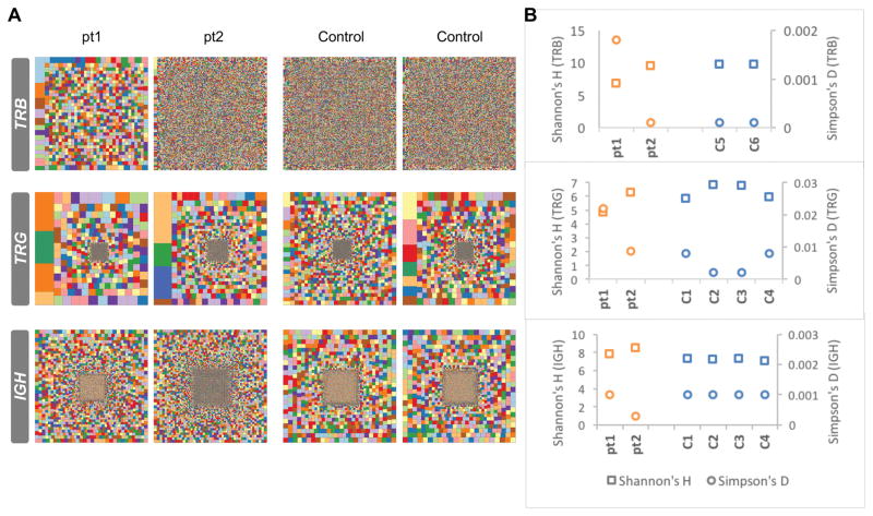
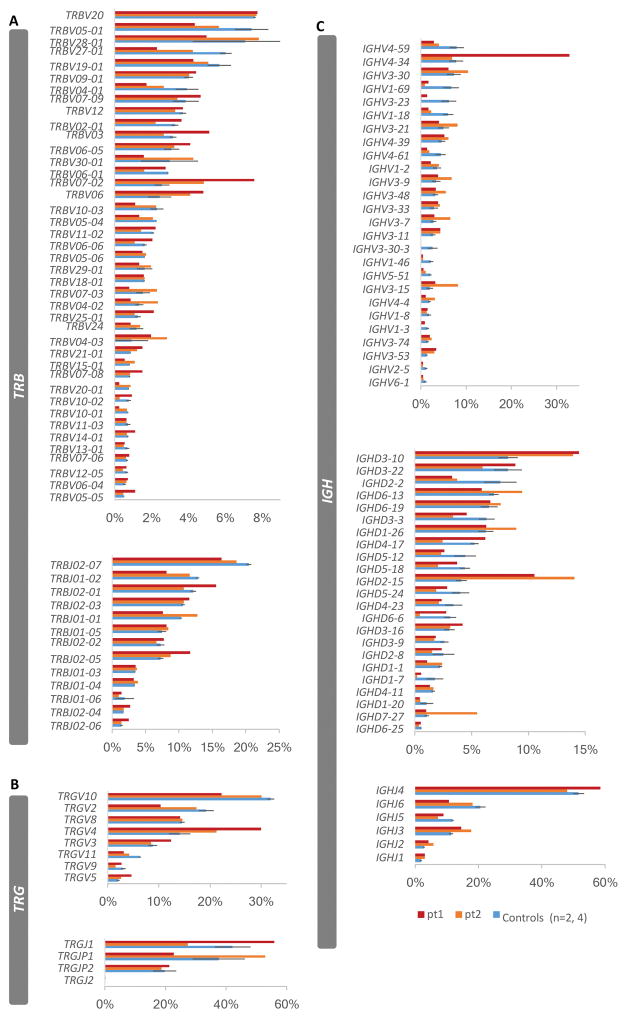
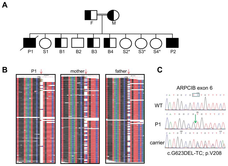
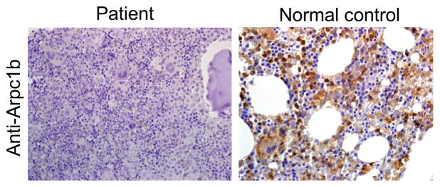
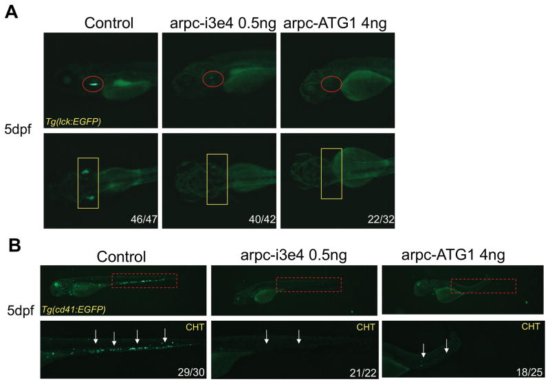
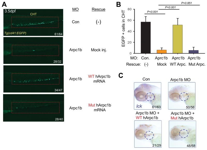
Regulation of the actin cytoskeleton is crucial for normal development and function of the immune system, as evidenced by the severe immune abnormalities exhibited by patients bearing inactivating mutations in the Wiskott-Aldrich syndrome protein (WASP), a key regulator of actin dynamics. WASP exerts its effects on actin dynamics through a multisubunit complex termed Arp2/3. Despite the critical role played by Arp2/3 as an effector of WASP-mediated control over actin polymerization, mutations in protein components of the Arp2/3 complex had not previously been identified as a cause of immunodeficiency. Here, we describe two brothers with hematopoietic and immunologic symptoms reminiscent of Wiskott-Aldrich syndrome (WAS). However, these patients lacked mutations in any of the genes previously associated with WAS. Whole-exome sequencing revealed a homozygous 2 bp deletion, n.c.G623DEL-TC (p.V208VfsX20), in Arp2/3 complex component ARPC1B that causes a frame shift resulting in premature termination. Modeling of the disease in zebrafish revealed that ARPC1B plays a critical role in supporting T cell and thrombocyte development. Moreover, the defects in development caused by ARPC1B loss could be rescued by the intact human ARPC1B ortholog, but not by the p.V208VfsX20 variant identified in the patients. Moreover, we found that the expression of ARPC1B is restricted to hematopoietic cells, potentially explaining why a mutation in ARPC1B has now been observed as a cause of WAS, whereas mutations in other, more widely expressed, components of the Arp2/3 complex have not been observed.


Conflict of interest statement

Conflict-of-interest disclosure

The authors declare no competing financial interests.

Figures

FIGURE 1
FIGURE 1. Analysis of the patients’ TRB, TRG and IGH repertoires
Hierarchical Treemaps (A), Shannon’s H, and Simpson’s D diversity indices (B) were generated for TRB, TRG and IGH repertoires by analysis of NGS of samples from two patients and two healthy donor controls for TRB repertoire and four healthy donor controls for TRG and IGH repertoires.
FIGURE 2
FIGURE 2. Differential VDJ gene usage relative to total sequences in the that are patients’ TRB, TRG and IGH repertoires
The frequencies of gene usages based on total sequences for the patients for TRBV and TRBJ genes (A), TRGV and TRGJ genes (B) and IGHV, IGHD and IGHJ genes (C), were compared to the frequencies (average ± SEM) of gene usage of two controls in TRB repertoire and four controls in TRG and IGH repertoires.
FIGURE 3
FIGURE 3. Genetic and genomic analysis of the patient’s inheritance of the ARPB1C mutation
A, Family pedigree. Solid symbols represent the affected subjects P1 (diagonal line, deceased) and P2. Half solid symbols represent unaffected relatives, which are carriers for the mutation. Open symbols represent unaffected relatives, including three sisters, marked by asterisks, that did not undergo genetic analysis for the familial mutation but clinically are unaffected. F=father; M=mother; P=patient; S=sister; B=brother. B, Whole exome sequencing of patient 1 and his parents (trio). Deep sequences around the 2 bp deletion (red arrow) are shown, demonstrating the homozygosity of the patient and the heterozygosity of his parents. C, Dideoxy Sanger sequencing of the different ARPC1B genotypes detected in the studied pedigree. The deleted TC nucleotides are boxed and their position in the patient’s sequence is marked by a green arrow.
FIGURE 4
FIGURE 4. Expression of ARPB1C protein in the patient’s bone marrow cells
Bone marrow was immunostained with anti-ARPC1B, which revealed that immunoreactivity to ARPB1C was absent from the patient’s bone marrow cells (left panel), but was abundant in the normal control (right panel). The magnification of the images in 400X.
FIGURE 5
FIGURE 5. Impairment of T cell and thrombocyte development upon knockdown of arpc1b in zebrafish
A, T cell development was disrupted after injection of MO, which disrupt arpc1b splicing (arpc-i3e4) or suppress translation of arpc1b mRNA (arpc-ATG1). T cell development was assessed at 5dpf using Tg(lck:EGFP) transgenic embryos to mark T cell progenitors (lateral view, red circles; dorsal view, yellow rectangles). B, Development of CD41+ thrombocytes was impaired following arpc1b knockdown. The effect of MO knockdown (as above) on thrombocyte development was assessed at 5dpf (red rectangles and white arrows). The embryos were photographed from lateral views. CHT: caudal hematopoietic tissue. The numbers on the images refer to the fraction of embryos with the depicted phenotype.
FIGURE 6
FIGURE 6. Patient mutation in ARPC1B fails to rescue developmental anomalies resulting from Arpc1b knockdown
A, Overexpression of human WT but not mutant ARPC1B mRNA (150pg) partially rescued the thrombocyte development in 3.5dpf Tg(CD41:EGFP) embryos (lateral view, red rectangles) treated with arpc1b-i3e4 MO (Arpc1b). The numbers refer to the fractions of embryos exhibiting the depicted phenotypes. B, Quantitation of EGFP+ cell numbers in the CHT region as depicted in the red boxes of Fig. 6A as the mean of five embryos of each phenotype. T bar indicates the standard deviation. C, Overexpression ARPC1B in arpc1b morphant embryos using heat-shock inducible plasmids (150pg) encoding WT or mutant human ARPC1B. Developing T cells were identified by performing WISH analysis at 5dpf for using a probe for lck (blue circles, lateral view). The numbers refer to the fractions of embryos exhibiting the depicted phenotypes.

References

    1. Pizarro-Cerda J, Chorev DS, Geiger B, Cossart P. The Diverse Family of Arp2/3 Complexes. Trends Cell Biol. 2017;27:93–100. - PMC - PubMed
    1. Tyler JJ, Allwood EG, Ayscough KR. WASP family proteins, more than Arp2/3 activators. Biochem Soc Trans. 2016;44:1339–1345. - PMC - PubMed
    1. Swaney KF, Li R. Function and regulation of the Arp2/3 complex during cell migration in diverse environments. Curr Opin Cell Biol. 2016;42:63–72. - PMC - PubMed
    1. Derry JM, Ochs HD, Francke U. Isolation of a novel gene mutated in Wiskott-Aldrich syndrome. Cell. 1994;78:635–644. - PubMed
    1. Chandra S, Bronicki L, Nagaraj CB, Zhang K. WAS-Related Disorders. In: Pagon RA, Adam MP, Ardinger HH, Wallace SE, Amemiya A, Bean LJH, Bird TD, Ledbetter N, Mefford HC, Smith RJH, Stephens K, editors. GeneReviews(R) Seattle (WA): 1993. - PubMed

Publication types

MeSH terms