. 2018 Mar 1;314(3):H659-H668.
doi: 10.1152/ajpheart.00452.2017. Epub 2017 Nov 10.

Protective effects of the mechanistic target of rapamycin against excess iron and ferroptosis in cardiomyocytes

Affiliations

Protective effects of the mechanistic target of rapamycin against excess iron and ferroptosis in cardiomyocytes

Yuichi Baba et al. Am J Physiol Heart Circ Physiol. .

Abstract

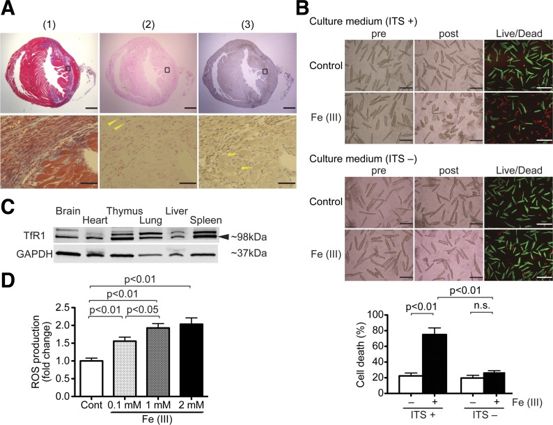
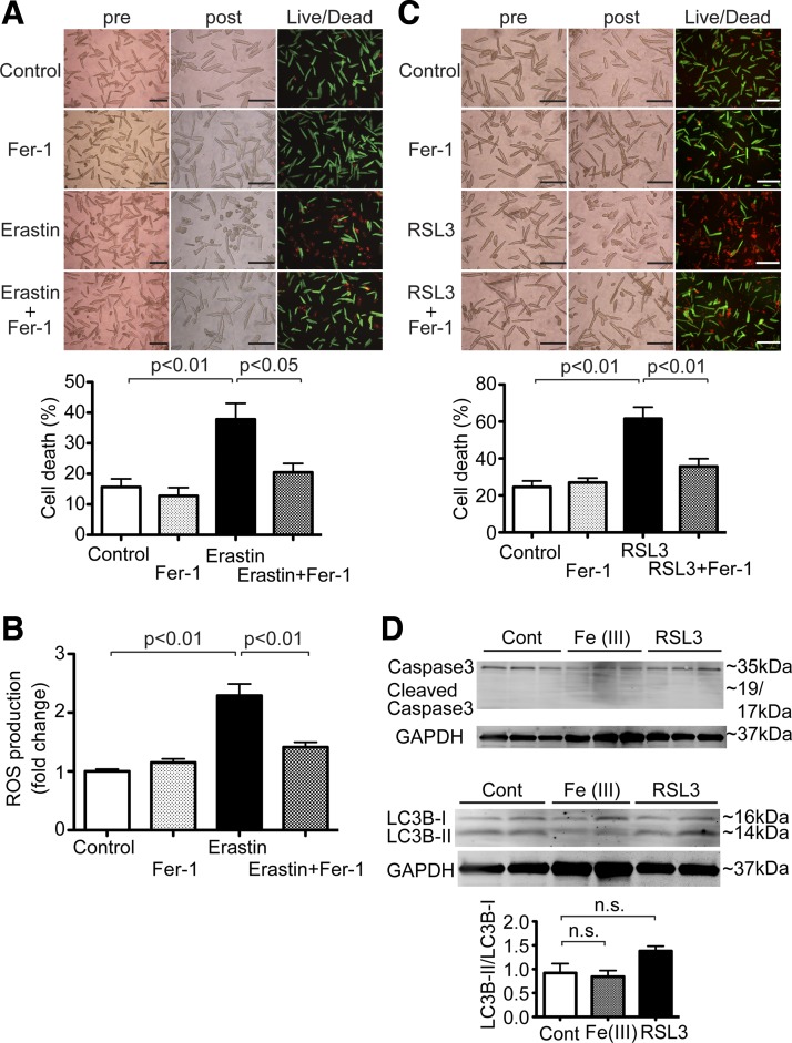
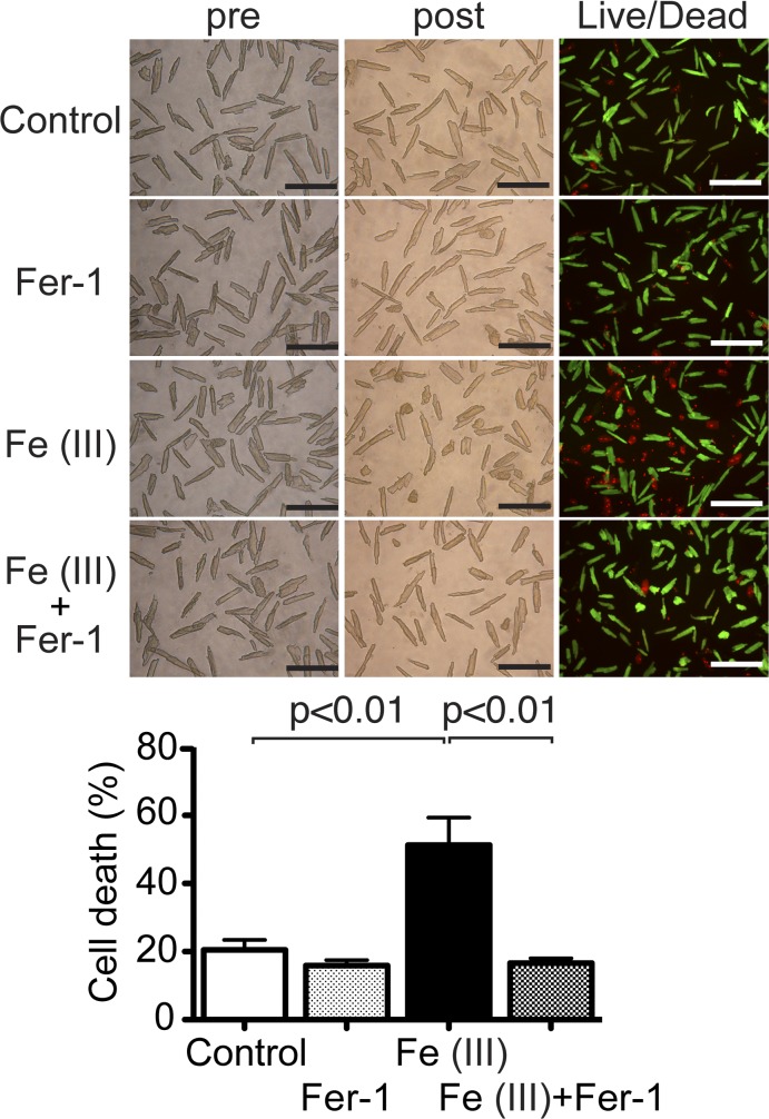
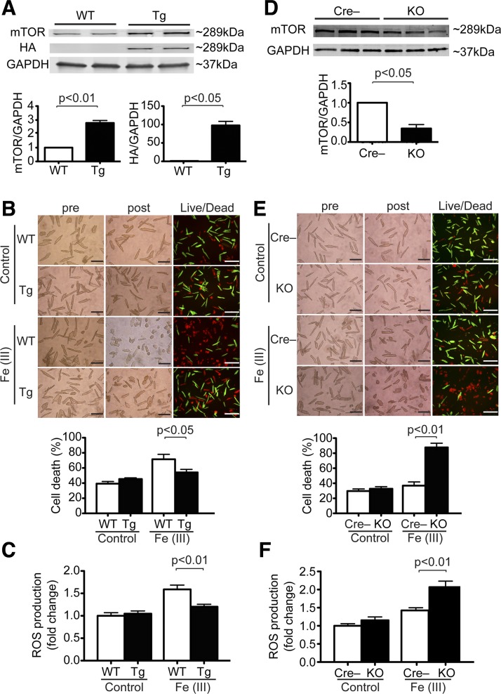
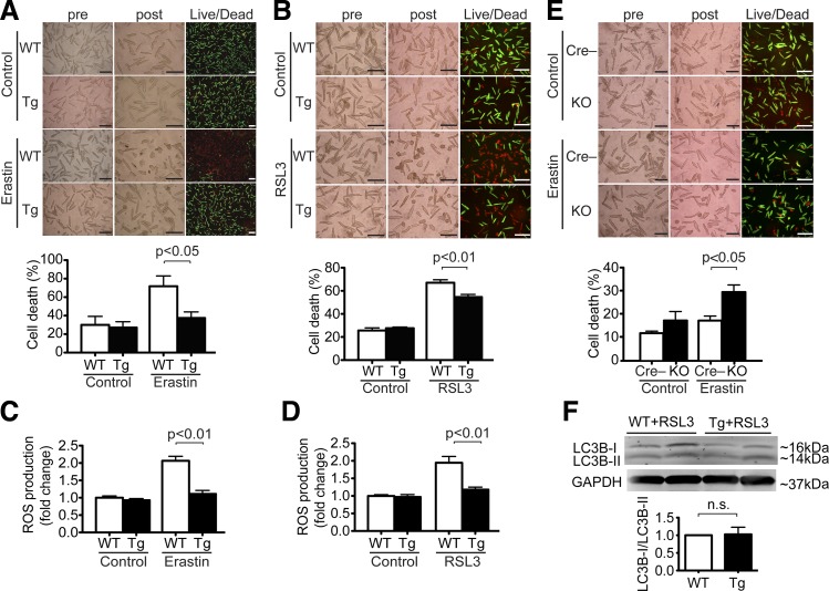
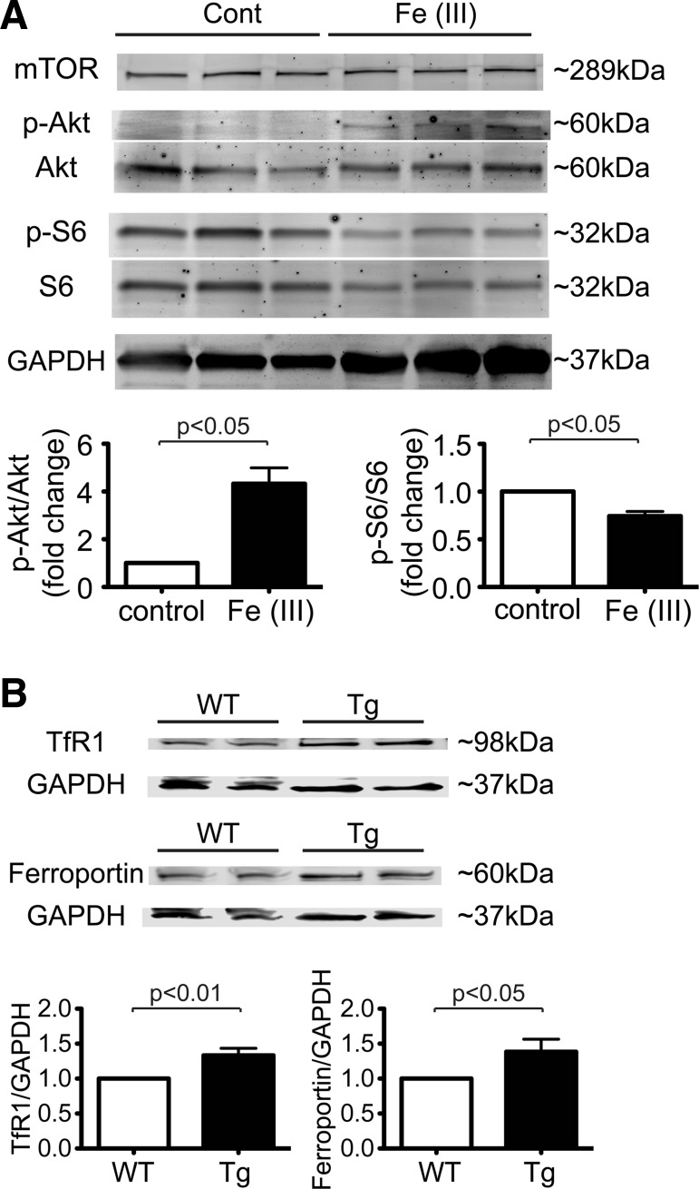
Clinical studies have suggested that myocardial iron is a risk factor for left ventricular remodeling in patients after myocardial infarction. Ferroptosis has recently been reported as a mechanism of iron-dependent nonapoptotic cell death. However, ferroptosis in the heart is not well understood. Mechanistic target of rapamycin (mTOR) protects the heart against pathological stimuli such as ischemia. To define the role of cardiac mTOR on cell survival in iron-mediated cell death, we examined cardiomyocyte (CM) cell viability under excess iron and ferroptosis conditions. Adult mouse CMs were isolated from cardiac-specific mTOR transgenic mice, cardiac-specific mTOR knockout mice, or control mice. CMs were treated with ferric iron [Fe(III)]-citrate, erastin, a class 1 ferroptosis inducer, or Ras-selective lethal 3 (RSL3), a class 2 ferroptosis inducer. Live/dead cell viability assays revealed that Fe(III)-citrate, erastin, and RSL3 induced cell death. Cotreatment with ferrostatin-1, a ferroptosis inhibitor, inhibited cell death in all conditions. mTOR overexpression suppressed Fe(III)-citrate, erastin, and RSL3-induced cell death, whereas mTOR deletion exaggerated cell death in these conditions. 2',7'-Dichlorodihydrofluorescein diacetate measurement of reactive oxygen species (ROS) production showed that erastin-induced ROS production was significantly lower in mTOR transgenic versus control CMs. These findings suggest that ferroptosis is a significant type of cell death in CMs and that mTOR plays an important role in protecting CMs against excess iron and ferroptosis, at least in part, by regulating ROS production. Understanding the effects of mTOR in preventing iron-mediated cell death will provide a new therapy for patients with myocardial infarction. NEW & NOTEWORTHY Ferroptosis has recently been reported as a new form of iron-dependent nonapoptotic cell death. However, ferroptosis in the heart is not well characterized. Using cultured adult mouse cardiomyocytes, we demonstrated that the mechanistic target of rapamycin plays an important role in protecting cardiomyocytes against excess iron and ferroptosis.

Keywords: cardiomyocyte; ferroptosis; iron; mechanistic target of rapamycin.


Figures

Fig. 1.
Fig. 1.
Excess iron induces cardiomyocyte (CM) cell death. A: iron and ferritin accumulation in the mouse heart after ischemia-reperfusion (I/R) injury. This is a representative sample from 6 mice at 1 wk post-I/R injury. All samples displayed a similar staining pattern to the one shown here. Top, Masson’s trichrome staining (1), fibrotic areas stained with blue (2), and iron staining (3). Anti-ferritin-positive cells are stained with brown (anti-ferritin staining). Scale bars = 1 mm. Bottom, magnified images of regions indicated by squares in each heart section of the top images (1), Masson’s trichrome staining and iron staining (2), and free iron-positive cells stained with blue (3); arrowheads indicate iron-positive non-CMs. Scale bars = 50 µm. B: excess iron-induced CM cell death. Top, representative images of CM cell death induced by Fe(III)-citrate in the presence or absence of insulin-transferrin-sodium selenite (ITS) in culture medium. Adult mouse CMs isolated from wild-type (WT) mice were exposed to 1 mM Fe(III)-citrate or 1 mM sodium citrate for 24 h. Morphological changes in the CM cell culture were assessed before applying the agents (pre) and at the end of experiments (post). Live/dead assays, in which live cells stain with calcein AM (green) and nuclei of dead cells stain with ethidium homodimer-1 (red), were used to assess cell viability. This is a set of representative images among 4 independent experiments. Scale bars = 200 µm. Bottom, quantitative data of CM cell death. Total numbers of live cytosol (green) and dead nuclei (red) cells were counted in three low-power fields. More than 500 cells were counted in each condition. The percent cell death was calculated as dead cells/total cells; n = 4. C: representative immunoblots of transferrin receptor 1 (TfR1) expression in mouse organs. Arrow indicates immunoblots of TfR1. Blots are representative from 3 independent WT mice. D: reactive oxygen species (ROS) production after Fe(III)-citrate treatment in WT CMs. ROS production in WT CMs exposed to 0.1, 1, or 2 mM Fe(III)-citrate or control buffer (Cont) for 24 h was measured by 2′,7′-dichlorodihydrofluorescein diacetate; n = 6. n.s., Not significant.
Fig. 2.
Fig. 2.
Ferroptosis in cardiomyocytes (CMs). A: erastin-induced CM cell death. Top, representative images of CM cell death induced by erastin. Adult mouse CMs isolated from wild-type (WT) mice were treated with 50 μM erastin or vehicle control in the presence or absence of 10 μM ferrostatin-1 (Fer-1) for 16 h. Morphological changes in CM cell culture were assessed before applying the agents (pre) and at the end of experiments (post). Cell death was assessed by a Live/Dead assay. Representative images are shown from 4 independent experiments. Scale bars = 200 µm. Bottom, quantitative data of CM cell death. Percent cell death was calculated as described in Fig. 1; n = 4. B: reactive oxygen species (ROS) production after erastin treatment in WT CMs. ROS production was assessed in WT CMs treated with 50 μM erastin or vehicle control in the presence or absence of 10 μM Fer-1 for 16 h. ROS production was measured by 2′,7′-dichlorodihydrofluorescein diacetate; n = 5. C: Ras-selective lethal 3 (RSL3)-induced CM cell death. Top, representative images of CM cell death induced by RSL3. Adult mouse CMs isolated from WT mice were treated with 1 μg/ml RSL3 or vehicle control in the presence or absence of 10 μM Fer-1 for 16 h. Representative images are shown from 6 independent experiments. Scale bars = 200 µm. Bottom, quantitative data of CM cell death. Percent cell death was calculated as described in Fig. 1; n = 6. D: expression levels of cleaved caspase-3 and light chain (LC)3B in WT CMs treated with vehicle control (Cont), 1 mM Fe(III)-citrate, or 1 μg/ml RSL3 for 16 h. Top, representative immunoblots of cleaved caspase-3 and LC3B in CMs. Bottom, densitometric quantitation of LC3B expression. Data are ratios of LC3B-II to LC3B-I. Control, n = 3; Fe(III), n = 3; RSL3, n = 3.
Fig. 3.
Fig. 3.
Ferrostatin-1 (Fer-1), a lipid reactive oxygen species (ROS) scavenger, inhibits excess iron-induced cell death in cardiomyocytes (CMs). Top, representative images of CM cell death induced by excess iron. Isolated CMs from wild-type (WT) adult mice were exposed to 1 mM Fe(III)-citrate or vehicle control for 24 h. To test the contribution of lipid ROS to iron-induced cell death, CMs in each group were cotreated with or without 10 μM Fer-1. Morphological changes in CM cell culture were assessed before applying agents (pre) and at the end of experiments (post). Cell death was assessed by Live/Dead assay. Representative images are shown from 5 independent experiments. Scale bars = 200 µm. Bottom, quantitative data of CM cell death. Percent cell death was calculated as described in Fig. 1; n = 5.
Fig. 4.
Fig. 4.
Mechanistic target of rapamycin (mTOR) protects against excess iron-induced cardiomyocyte (CM) cell death. A: expression levels of mTOR and hemagglutinin (HA) in CMs isolated from wild-type (WT) and mTOR transgenic (Tg) mice. Top, representative immunoblots of mTOR and HA in CMs. Blots are representative from 3 WT mice and 3 Tg mice. Bottom, densitometric quantitation of mTOR and HA expression. Data were normalized to GAPDH. WT, n = 3; Tg, n = 3. B: effects of excess iron on CMs from Tg mice. Adult CMs isolated from either WT or Tg mice were exposed to 2 mM Fe(III)-citrate or vehicle control for 24 h. Top, morphological changes in CM cell culture were assessed before applying the agents (pre) and at the end of experiments (post). Cell death was assessed by Live/Dead assay. Representative images are shown from 6 WT mice and 6 Tg mice. Scale bars = 200 µm. Bottom, quantitative data of CM cell death. Percent cell death was calculated as described in Fig. 1. WT, n = 6; Tg, n = 6. C: ROS production in WT and Tg CMs treated with 1 mM Fe(III)-citrate or vehicle control for 8 h. ROS production was measured by 2′,7′-dichlorodihydrofluorescein diacetate (H2DCFDA). WT, n = 4; Tg, n = 4. D: expression levels of cardiac mTOR in mTOR knockout (KO) mice. Top, immunoblots of mTOR in CMs isolated from Cre-negative control (Cre) and mTOR KO mice. Blots are representative from 3 Cre mice and 3 KO mice. Bottom, densitometric quantitation of mTOR expression in CMs in KO mice. Data were normalized to GAPDH. Cre, n = 3; KO, n = 3. E: effects of excess iron in CMs from KO mice. Adult CMs isolated from either Cre or KO mice were exposed to 1 mM Fe(III)-citrate or vehicle control for 24 h. Top, morphological changes and Live/Dead assay. Representative images are shown from 3 Cre mice and 3 KO mice. Scale bars = 200 µm. Bottom, quantitative data of CM cell death. Percent cell death was calculated as described in Fig. 1. Cre, n = 3; KO, n = 3. F: ROS production in Cre and KO CMs treated with 1 mM Fe(III)-citrate or vehicle control for 8 h. ROS production was measured by H2DCFDA. Cre, n = 3; KO, n = 3.
Fig. 5.
Fig. 5.
Mechanistic target of rapamycin (mTOR) protects cardiomyocytes (CMs) against ferroptosis. A: effects of erastin in CMs from mTOR transgenic (Tg) mice. Adult CMs isolated from wild-type (WT) or Tg mice were treated with 40 μM erastin or vehicle control for 16 h in CMs. Top, morphological changes in CM cell culture were assessed before applying agents (pre) and at the end of experiments (post). Cell death was assessed by Live/Dead assay. Representative images are shown from 5 WT mice and 4 Tg mice. Scale bars = 200 µm. Bottom, quantitative data of CM cell death. Percent cell death was calculated as described in Fig. 1. WT, n = 5; Tg, n = 4. B: effects of Ras-selective lethal 3 (RSL3) in CMs from Tg mice. Adult CMs isolated from WT or Tg mice were treated with 1 μg/ml RSL3 or vehicle control for 16 h in CMs. Top, morphological changes and Live/Dead assay. Representative images are shown from 6 WT mice and 6 Tg mice. Scale bars = 200 µm. Bottom, quantitative data of CM cell death. Percent cell death was calculated as described in Fig. 1. WT, n = 6; Tg, n = 6. C: reactive oxygen species (ROS) production in WT and Tg CMs treated with 40 μM erastin or vehicle control for 8 h. ROS production was measured by 2′,7′-dichlorodihydrofluorescein diacetate (H2DCFDA). WT, n = 6; Tg, n = 6. D: ROS production in WT and Tg CMs treated with 1 μg/ml RSL3 or vehicle control for 8 h. ROS production was measured by H2DCFDA. WT, n = 3; Tg, n = 3. E: effects of erastin in CMs from mTOR knockout (KO) mice. Adult CMs isolated Cre-negative control (Cre) and KO mice were treated with 20 μM erastin or vehicle control for 16 h in CMs. Top, morphological changes and Live/Dead assay. Representative images are shown from 6 Cre mice and 6 KO mice. Scale bars = 200 µm. Bottom, quantitative data of CM cell death. Percent cell death was calculated as described in Fig. 1. Cre, n = 6; KO, n = 6. F: expression levels of light chain (LC)3B in CMs from WT and Tg mice treated with 1 μg/ml RSL3 for 16 h. Top, representative immunoblots of LC3B in CMs. Bottom, densitometric quantitation of LC3B expression. Data are ratios of LC3B-II to LC3B-I. WT, n = 3; Tg, n = 3.
Fig. 6.
Fig. 6.
Mechanistic target of rapamycin (mTOR) increases the expression levels of transferrin receptor 1 (TfR1) and ferroportin. A, top: representative immunoblots of mTOR signaling in cardiomyocytes (CMs). CMs isolated from wild-type (WT) mice were treated with 1 mM Fe(III)-citrate or vehicle control (Control) for 16 h. Immunoblot analysis was performed with the indicated antibodies. Bottom, densitometric quantitative analyses of phosphorylated (p-)Akt and p-S6 are shown with the ratio of p-Akt to total Akt (Akt) and the ratio of p-S6 to total S6 (S6). Data were normalized to those of control cells. Control, n = 3; Fe(III), n = 3. B, top: representative immunoblots of TfR1 and ferroportin in hearts harvested from WT and mTOR transgenic (Tg) mice without additional treatments. Blots are representative of 6 WT mice and 6 Tg mice for TfR1 and 9 WT mice and 9 Tg mice for ferroportin. Bottom, densitometric quantitation of TfR1 and ferroportin expression. Data were normalized to GAPDH. n = 6 WT mice and n = 6 mice for TfR1; n = 9 WT mice and n = 9 Tg mice for ferroportin.

Comment in

  • Ferroptosis: beating on death's door.
    Dhingra R, Ravandi A, Kirshenbaum LA. Dhingra R, et al. Am J Physiol Heart Circ Physiol. 2018 Apr 1;314(4):H772-H775. doi: 10.1152/ajpheart.00692.2017. Epub 2017 Dec 6. Am J Physiol Heart Circ Physiol. 2018. PMID: 29212794 No abstract available.

References

    1. Aoyagi T, Higa JK, Aoyagi H, Yorichika N, Shimada BK, Matsui T. Cardiac mTOR rescues the detrimental effects of diet-induced obesity in the heart after ischemia-reperfusion. Am J Physiol Heart Circ Physiol 308: H1530–H1539, 2015. doi:10.1152/ajpheart.00008.2015. - DOI - PMC - PubMed
    1. Aoyagi T, Kusakari Y, Xiao CY, Inouye BT, Takahashi M, Scherrer-Crosbie M, Rosenzweig A, Hara K, Matsui T. Cardiac mTOR protects the heart against ischemia-reperfusion injury. Am J Physiol Heart Circ Physiol 303: H75–H85, 2012. doi:10.1152/ajpheart.00241.2012. - DOI - PMC - PubMed
    1. Aoyagi T, Matsui T. Phosphoinositide-3 kinase signaling in cardiac hypertrophy and heart failure. Curr Pharm Des 17: 1818–1824, 2011. doi:10.2174/138161211796390976. - DOI - PMC - PubMed
    1. Barth S, Glick D, Macleod KF. Autophagy: assays and artifacts. J Pathol 221: 117–124, 2010. doi:10.1002/path.2694. - DOI - PMC - PubMed
    1. Bayeva M, Khechaduri A, Puig S, Chang HC, Patial S, Blackshear PJ, Ardehali H. mTOR regulates cellular iron homeostasis through tristetraprolin. Cell Metab 16: 645–657, 2012. doi:10.1016/j.cmet.2012.10.001. - DOI - PMC - PubMed

Publication types

MeSH terms