

Human Gingiva-Derived Mesenchymal Stem Cells Ameliorate Streptozoticin-induced T1DM in mice via Suppression of T effector cells and Up-regulating Treg Subsets
- PMID: 29127315
- PMCID: PMC5681565
- DOI: 10.1038/s41598-017-14979-5
Human Gingiva-Derived Mesenchymal Stem Cells Ameliorate Streptozoticin-induced T1DM in mice via Suppression of T effector cells and Up-regulating Treg Subsets
Abstract
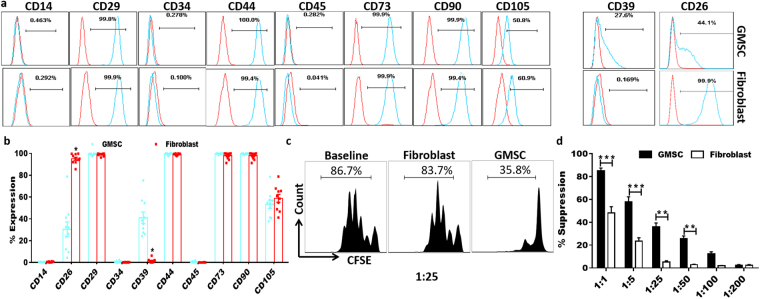
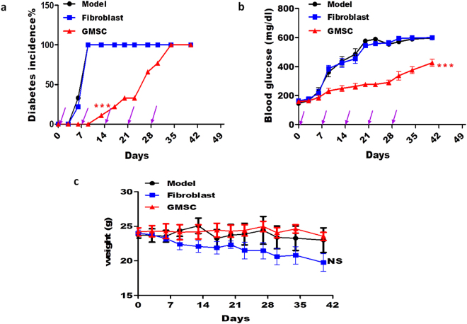
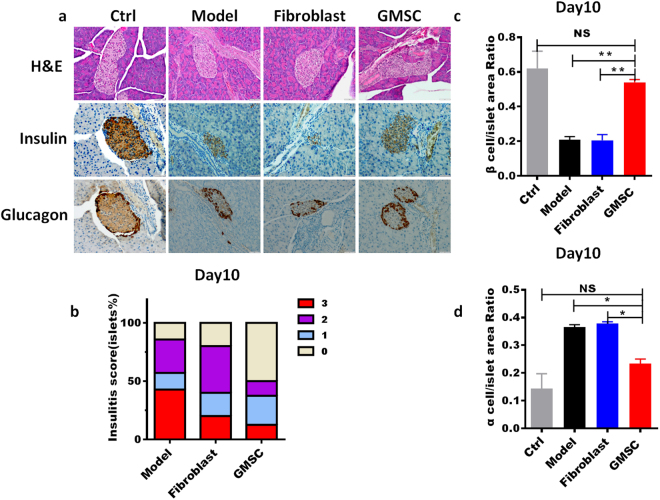
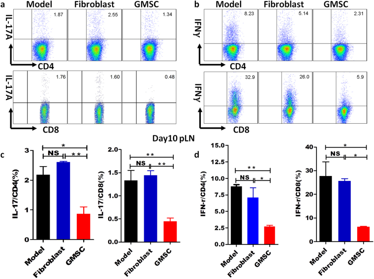
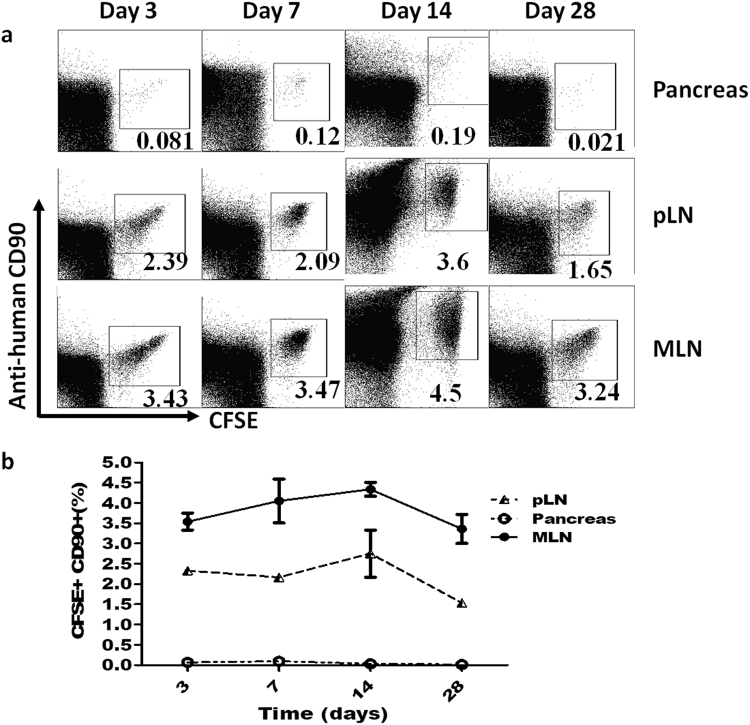
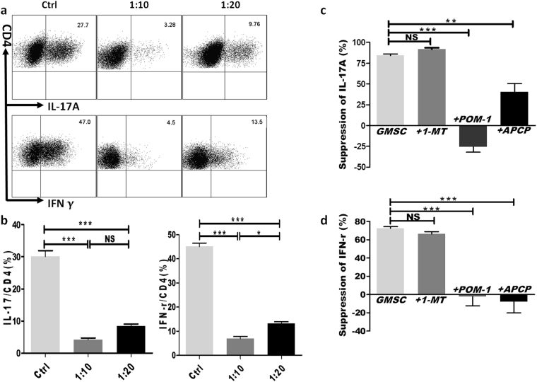
There is yet no cure for type 1 diabetes (T1DM) so far. A significant body of evidence has demonstrated that bone marrow-derived mesenchymal stem cells (BMSCs) showed great potential in controlling T1DM. But there exists much difficulty in using BMSCs as a clinical therapy. We here test whether a new population of mesenchymal stem cells from human gingiva (GMSCs), which has many advantages over BMSCs, can delay or prevent progress of T1DM. GMSCs were adoptively transferred to multiple low-dose streptozotocin (STZ)-induced T1DM. Blood glucose levels and disease severities were analyzed. T cells subsets in blood, spleen and lymph nodes were detected dynamically by flow cytometry. GMSC distribution was dynamically analyzed. We found that infusion of GMSCs but not fibroblast cells significantly controlled blood glucose levels, delayed diabetes onset, ameliorated pathology scores in pancreas, and down-regulated production of IL-17 and IFN-γ in CD4+ and CD8+ T cells in spleens, pancreatic lymph nodes (pLN) and other lymph nodes. GMSCs also up-regulated the levels of CD4+ Treg induced in the periphery. Mechanismly, GMSCs could migrate to pancreas and local lymph node and function through CD39/CD73 pathway to regulate effector T cells. Thus, GMSCs show a potential promise in treating T1DM in the clinic.
Conflict of interest statement
The authors declare that they have no competing interests.
Figures

Similar articles
-
Human gingiva-derived mesenchymal stem cells alleviate inflammatory bowel disease via IL-10 signalling-dependent modulation of immune cells.Scand J Immunol. 2019 Sep;90(3):e12751. doi: 10.1111/sji.12751. Epub 2019 Jul 8. Scand J Immunol. 2019. PMID: 30664802
-
Transplantation of gingiva-derived mesenchymal stem cells ameliorates collagen-induced arthritis.Arthritis Res Ther. 2016 Nov 11;18(1):262. doi: 10.1186/s13075-016-1160-5. Arthritis Res Ther. 2016. PMID: 27836015 Free PMC article.
-
[Acetylsalicylic acid treatment enhanced immunomodulatory function of mesenchymal stem cells derived from gingiva].Beijing Da Xue Xue Bao Yi Xue Ban. 2017 Oct 18;49(5):872-877. Beijing Da Xue Xue Bao Yi Xue Ban. 2017. PMID: 29045972 Chinese.
-
Updates on GMSCs Treatment for Autoimmune Diseases.Curr Stem Cell Res Ther. 2018;13(5):345-349. doi: 10.2174/1574888X13666180220141114. Curr Stem Cell Res Ther. 2018. PMID: 29468980 Review.
-
Stemness Potency of Human Gingival Cells-Application in Anticancer Therapies and Clinical Trials.Cells. 2020 Aug 18;9(8):1916. doi: 10.3390/cells9081916. Cells. 2020. PMID: 32824702 Free PMC article. Review.
Cited by
-
Advanced Delivery Strategies for Immunotherapy in Type I Diabetes Mellitus.BioDrugs. 2023 May;37(3):331-352. doi: 10.1007/s40259-023-00594-6. Epub 2023 May 13. BioDrugs. 2023. PMID: 37178431 Free PMC article. Review.
-
Mesenchymal Stem Cell-Derived Exosomes: A Promising Biological Tool in Nanomedicine.Front Pharmacol. 2021 Jan 25;11:590470. doi: 10.3389/fphar.2020.590470. eCollection 2020. Front Pharmacol. 2021. PMID: 33716723 Free PMC article. Review.
-
Gingival mesenchymal stem cells: Biological properties and therapeutic applications.J Oral Biol Craniofac Res. 2024 Sep-Oct;14(5):547-569. doi: 10.1016/j.jobcr.2024.07.003. Epub 2024 Jul 13. J Oral Biol Craniofac Res. 2024. PMID: 39108352 Free PMC article.
-
Effect and mechanism of propranolol on promoting osteogenic differentiation and early implant osseointegration.Int J Mol Med. 2021 Oct;48(4):191. doi: 10.3892/ijmm.2021.5024. Epub 2021 Aug 20. Int J Mol Med. 2021. PMID: 34414453 Free PMC article.
-
Role and application of stem cells in dental regeneration: A comprehensive overview.EXCLI J. 2021 Feb 22;20:454-489. doi: 10.17179/excli2021-3335. eCollection 2021. EXCLI J. 2021. PMID: 33746673 Free PMC article. Review.
References
-
- Alberti KG, Zimmet PZ. Definition, diagnosis and classification of diabetes mellitus and its complications. Part 1: diagnosis and classification of diabetes mellitus provisional report of a WHO consultation. Diabet Med. 1998;15:539–553. doi: 10.1002/(SICI)1096-9136(199807)15:7<539::AID-DIA668>3.0.CO;2-S. - DOI - PubMed
Publication types
MeSH terms
Grants and funding
LinkOut - more resources
Full Text Sources
Other Literature Sources
Medical
Research Materials
