. 2017 Nov 10;7(1):15300.
doi: 10.1038/s41598-017-15563-7.

Catabolic Effects of Human PTH (1-34) on Bone: Requirement of Monocyte Chemoattractant Protein-1 in Murine Model of Hyperparathyroidism

Affiliations

Catabolic Effects of Human PTH (1-34) on Bone: Requirement of Monocyte Chemoattractant Protein-1 in Murine Model of Hyperparathyroidism

Jawed A Siddiqui et al. Sci Rep. .

Abstract

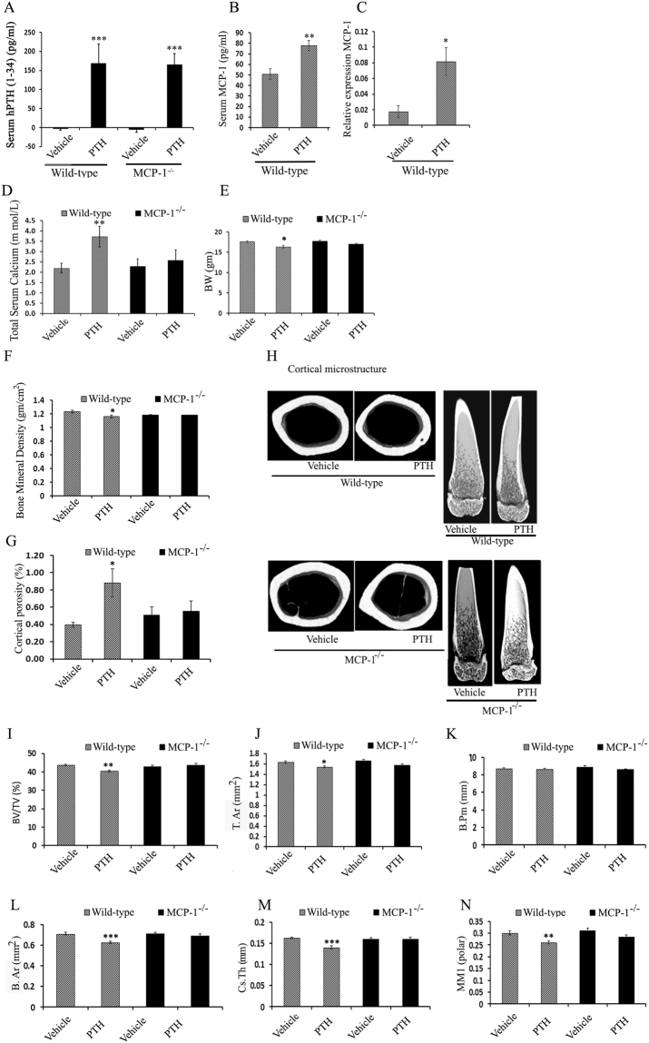
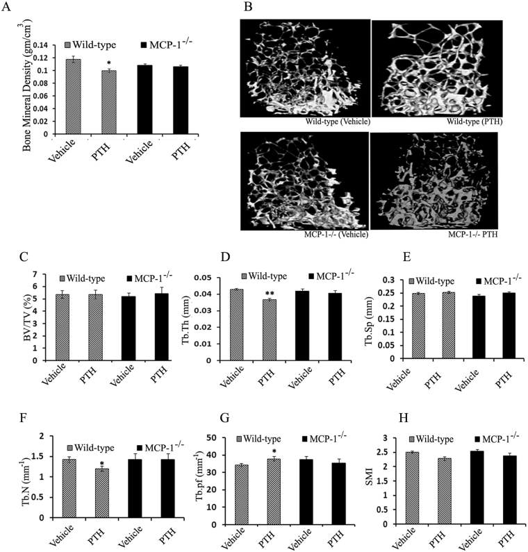
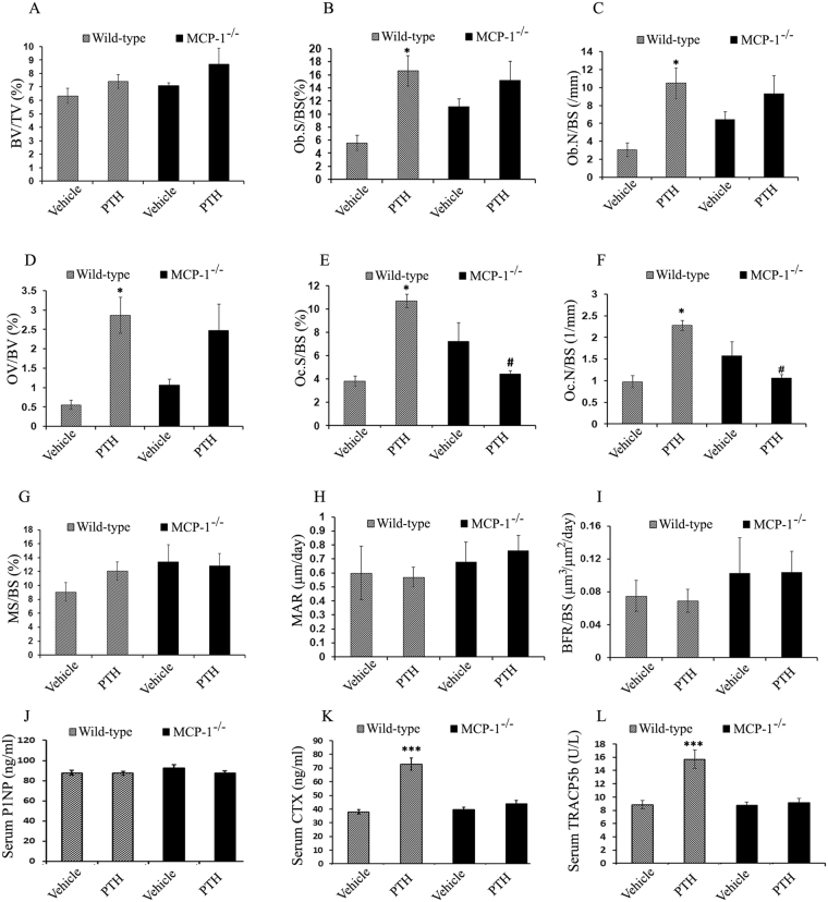
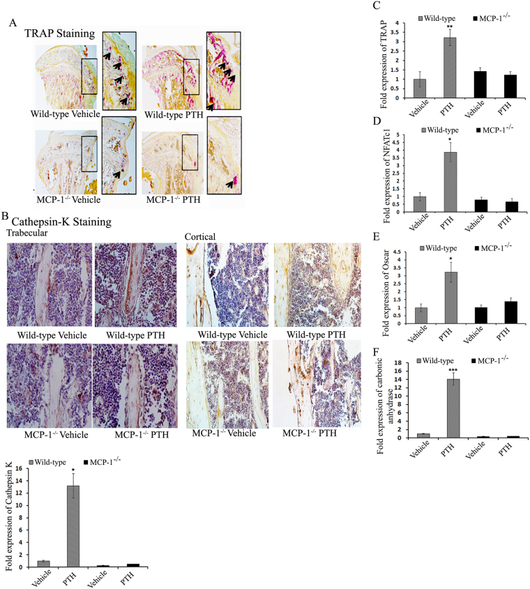
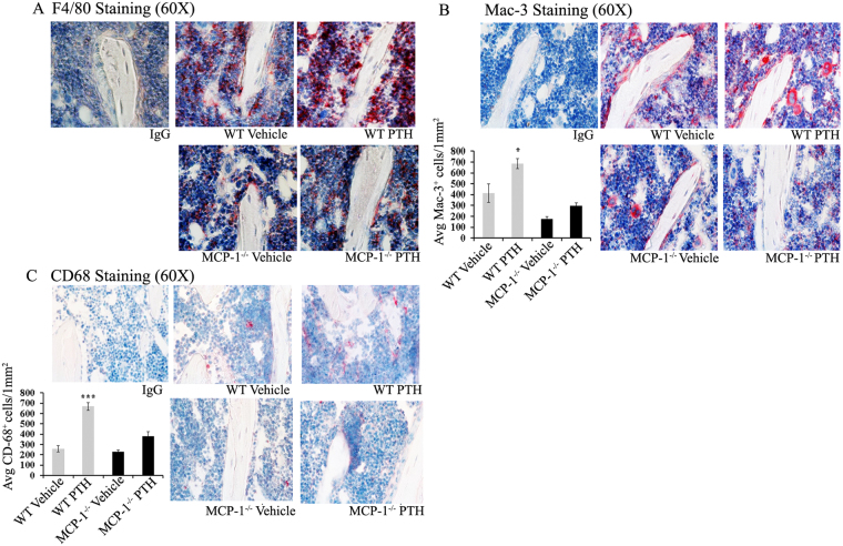
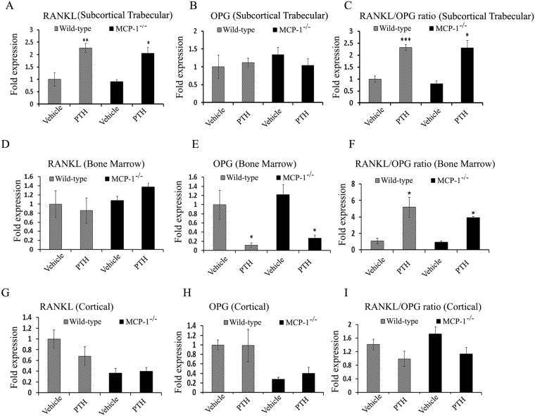
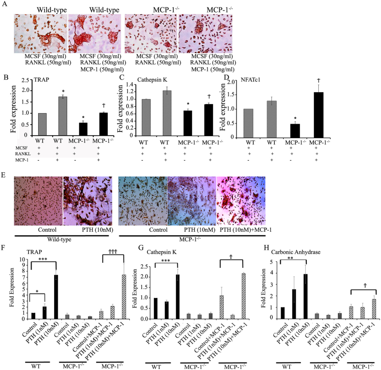
The bone catabolic actions of parathyroid hormone (PTH) are seen in patients with hyperparathyroidism, or with infusion of PTH in rodents. We have previously shown that the chemokine, monocyte chemoattractant protein-1 (MCP-1), is a mediator of PTH's anabolic effects on bone. To determine its role in PTH's catabolic effects, we continuously infused female wild-type (WT) and MCP-1-/- mice with hPTH or vehicle. Microcomputed tomography (µCT) analysis of cortical bone showed that hPTH-infusion induced significant bone loss in WT mice. Further, μCT analysis of trabecular bone revealed that, compared with the vehicle-treated group, the PTH-treated WT mice had reduced trabecular thickness and trabecular number. Notably, MCP-1-/- mice were protected against PTH-induced cortical and trabecular bone loss as well as from increases in serum CTX (C-terminal crosslinking telopeptide of type I collagen) and TRACP-5b (tartrate-resistant acid phosphatase 5b). In vitro, bone marrow macrophages (BMMs) from MCP-1-/- and WT mice were cultured with M-CSF, RANKL and/or MCP-1. BMMs from MCP-1-/- mice showed decreased multinucleated osteoclast formation compared with WT mice. Taken together, our work demonstrates that MCP-1 has a role in PTH's catabolic effects on bone including monocyte and macrophage recruitment, osteoclast formation, bone resorption, and cortical and trabecular bone loss.


Conflict of interest statement

The authors declare that they have no competing interests.

Figures

Figure 1
Figure 1
MCP-1 is required for cPTH-induced cortical bone loss. (A) Effect of continuous hPTH infusion on serum hPTH levels. Serum hPTH significantly increased in WT and MCP-1−/− mice. (B) Serum MCP-1 levels are significantly increased in WT mice after cPTH infusion. (C) MCP-1 gene expression after cPTH infusion. (D) Total serum calcium (mmol/L). (E) Change in body weight (BW) in gram (gm). (F) Cortical bone mineral density (vBMD cortical) in (gm/cm3). MCP-1−/− mice did not show loss of cortical BMD after cPTH infusion as seen in WT mice. (G) cPTH infusion increased cortical porosity in WT mice while not affecting this parameter in MCP-1−/− mice. (H) Representative µCT images of the cortical region of the femurs. (I–N) µCT analysis of cortical bone in the femurs of mice showing BV/TV (%), T.Ar (mm2), B.Pm (mm), B.Ar (mm2), Cs.Th (mm) and MMI polar. All values are expressed as mean ± SEM (n = 10–14 mice/group) *p < 0.05, **p < 0.01 and ***p < 0.001 compared to vehicle-infused WT mice.
Figure 2
Figure 2
MCP-1 null mice are protected from cPTH-induced trabecular bone loss. (A) Trabecular bone mineral density (vBMD trabecular) in (gm/cm3). (B) Representative µCT images of the trabecular region of femurs. (C–H) µCT analysis of trabecular bone in the femurs showing BV/TV (%), Tb.Th (mm), Tb.Sp (mm), Tb.N (1/mm), Tb.Pf (1/mm) and SMI. All values are expressed as mean ± SEM (n = 10–14 mice/group). *p < 0.05 and **p < 0.01 compared to vehicle-infused WT mice.
Figure 3
Figure 3
Histomorphometric analysis of femurs of mice (WT and MCP-1−/−) infused with cPTH for 2 weeks and biochemical markers. (A–F) Static histomorphometric analysis of femurs of WT and MCP-1−/− mice revealed a significant increase in osteoblast surface (Ob.S), osteoblast number (Ob.N), osteoid surface (OV/BV), osteoclast surface (Oc.S) and osteoclast number (Oc.N) in WT mice after 2 weeks of cPTH infusion. In contrast, cPTH-stimulated osteoblast and osteoclast activities are abolished in MCP-1 null mice. (G–I) There are no significant changes in analyzed dynamic histomorphometric parameters. (n = 6 mice/group). (J) The levels of P1NP, a marker of bone formation is unchanged in all groups; (K) CTX, a marker of bone resorption, (L) TRACP5b (surrogate for osteoclast number), a marker of bone resorption are increased with cPTH in WT mice but not in MCP-1−/− mice. All values are expressed as mean ± SEM (n = 10–14 mice/group). *p < 0.05 and ***p < 0.001 compared to vehicle-infused WT mice. #p < 0.05 compared to PTH-infused WT mice.
Figure 4
Figure 4
MCP-1 is necessary for cPTH-mediated osteoclastogenesis and osteoclast activity. (A) TRAP staining of tibiae from WT and MCP-1−/− mice (magnification 20X) shows less osteoclasts in MCP-1−/− mice with or without cPTH. (B) Immunohistochemistry for cathepsin K (known to be a more specific marker of osteoclasts) confirms the observations in (A). Lower panel, cathepsin K mRNA expression after PTH infusion. (C–F) mRNAs extracted from distal femur metaphyses and RT-qPCR was performed to examine the expression of TRAP, NFATc1, Oscar and carbonic anhydrase. No increase in any of these genes was seen in the MCP-1−/− mice infused with PTH in contrast to the WT mice. *p < 0.05, **p < 0.01, ***p < 0.001 compared with vehicle-infused WT mice.
Figure 5
Figure 5
Continuous PTH recruits increased numbers of macrophages in bone marrow and this is ablated in MCP-1−/− mice. (A) Two weeks of cPTH infusion increased the osteomac population in WT mice compared to vehicle-infused WT mice. MCP-1−/− mice showed fewer F4/80 positive cells in both vehicle and PTH-infused groups. Immunohistochemistry of tibial sections with F4/80 (magnification 60X). (B) Sections of WT and MCP-1−/− mice immunostained for Mac-3 and quantitation of stained cells. (C) CD68 immunostained sections of tibiae of WT and MCP-1−/− mice infused with cPTH or vehicle and quantitation of stained cells. There is a modest expression of Mac-3 and CD68 in MCP-1 null mice, with and without cPTH infusion (magnification 60X) and no stimulation with PTH. IgG was used as negative control for each stained section. *p < 0.05, ***p < 0.001 compared to vehicle-infused WT mice.
Figure 6
Figure 6
MCP-1 regulates cPTH-induced bone loss independent of the RANKL/OPG axis. WT and MCP-1−/− mice were infused with cPTH for 2 weeks. Subcortical trabecular, bone marrow and cortical bone compartments were separated, and RT-qPCR was performed to determine the differential expression of RANKL and OPG in these different compartments. (A,B,C) Increased expression of RANKL in subcortical trabecular bone after cPTH in both WT and MCP-1−/− mice, (A) RANKL, (B), OPG, (C), RANKL/OPG ratio. (D,E,F) Decreased expression of OPG in bone marrow after cPTH in both WT and MCP-1−/− mice, (D) RANKL, (E), OPG, (F), RANKL/OPG ratio. (G,H,I) Expression in cortical bone, (G) RANKL, (H), OPG, (I), RANKL/OPG ratio. (n = 4 mice in each group) *p < 0.05, **p < 0.01, ***p < 0.001 compared to vehicle-infused WT or MCP-1−/− mice.
Figure 7
Figure 7
MCP-1 promotes osteoclastogenesis in vitro and MCP-1 null mice exhibited impaired PTH-mediated bone marrow osteoclast formation. (A) Bone marrow cells from WT or MCP-1−/− mice were induced to differentiate towards the osteoclast lineage with M-CSF and RANKL for 7 days in the presence or absence of exogenous MCP-1 (50 ng/ml). Cultures were stained for TRAP and photographed by light microscopy. (BD) RT-qPCR analyses showing mRNA expression of osteoclast markers (TRAP, Cathepsin K and NFATc1) using the same cells as in (A) (n = 3). *p < 0.05 compared to cells from WT mice treated with M-CSF and RANKL, p < 0.05 compared to MCP-1−/− BMMs treated with M-CSF and RANKL. (E) Bone marrows from WT and MCP-1−/− mice were cultured for 10 days in the presence or absence of hPTH(1–34; 10−9 or 10−8M) and/or MCP-1 (50 ng/ml) to induce osteoclast formation. Cultures were stained for TRAP to identify multinucleated TRAP-positive cells. Representative images by light microscopy (4X). (FH) Bone marrows from WT and MCP-1−/− mice were cultured for 10 days in the presence or absence of hPTH(1–34; 10−9 or 10−8M) and/or MCP-1 (50 ng/ml) to induce osteoclast formation. Isolated RNAs were used for RT-qPCR analysis of osteoclast specific gene expression (TRAP, cathepsin K and carbonic anhydrase). *p < 0.05, **p < 0.01, ***p < 0.001 compared to Control cells from WT mice and p < 0.05, †††p < 0.001 compared to Control + MCP-1 -treated cells from MCP-1−/− mice.

Similar articles

Cited by

References

    1. Nakayama K, et al. Differences in bone and vitamin D metabolism between primary hyperparathyroidism and malignancy-associated hypercalcemia. J. Clin. Endocrinol. Metab. 1996;81:607–611. - PubMed
    1. Tangirala RK, Murao K, Quehenberger O. Regulation of expression of the human monocyte chemotactic protein-1 receptor (hCCR2) by cytokines. J. Biol. Chem. 1997;272:8050–8056. doi: 10.1074/jbc.272.12.8050. - DOI - PubMed
    1. Hopwood B, Tsykin A, Findlay DM, Fazzalari NL. Gene expression profile of the bone microenvironment in human fragility fracture bone. Bone. 2009;44:87–101. doi: 10.1016/j.bone.2008.08.120. - DOI - PubMed
    1. Lu Y, et al. CCCR2 expression correlates with prostate cancer progression. J. Cell Biochem. 2007;101:676–685. doi: 10.1002/jcb.21220. - DOI - PubMed
    1. Lu Y, et al. Monocyte chemotactic protein-1 mediates prostate cancer-induced bone resorption. Cancer Res. 2007;67:3646–3653. doi: 10.1158/0008-5472.CAN-06-1210. - DOI - PubMed

Publication types

MeSH terms