

Ultrafiltration combined with size exclusion chromatography efficiently isolates extracellular vesicles from cell culture media for compositional and functional studies
- PMID: 29127410
- PMCID: PMC5681555
- DOI: 10.1038/s41598-017-15717-7
Ultrafiltration combined with size exclusion chromatography efficiently isolates extracellular vesicles from cell culture media for compositional and functional studies
Abstract
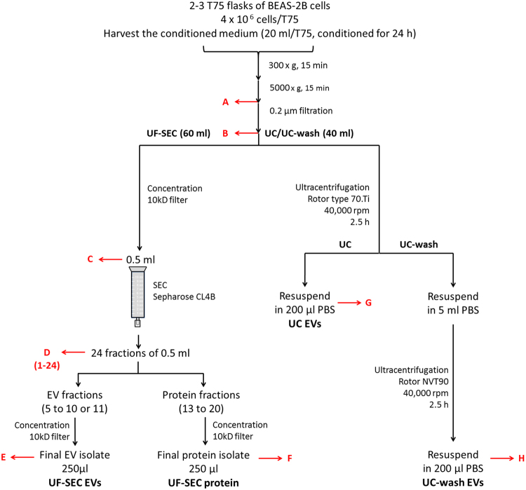
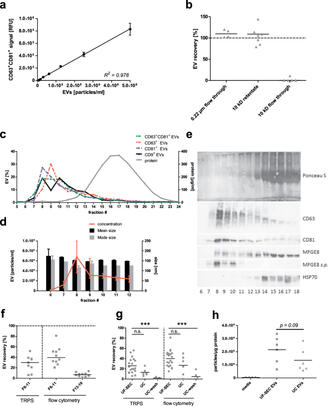
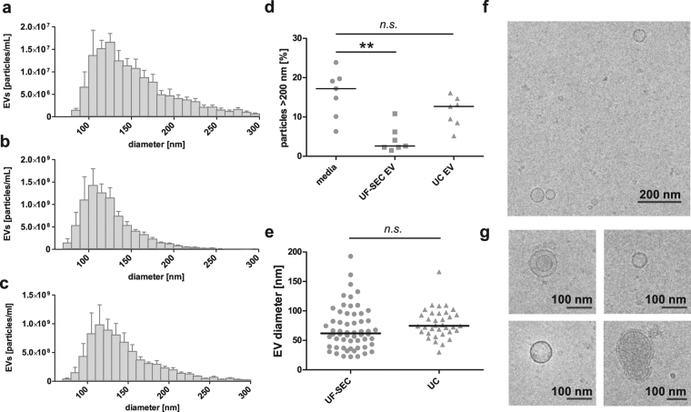
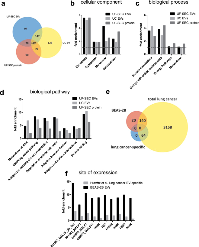
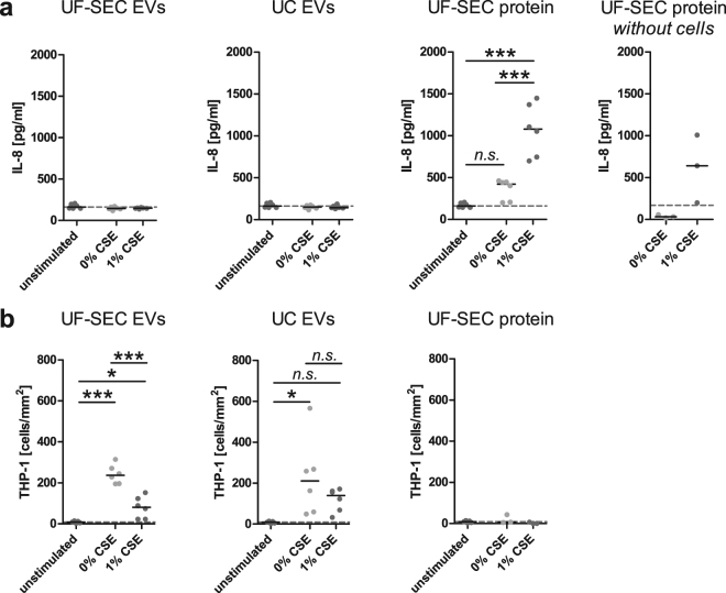
Appropriate isolation methods are essential for unravelling the relative contribution of extracellular vesicles (EVs) and the EV-free secretome to homeostasis and disease. We hypothesized that ultrafiltration followed by size exclusion chromatography (UF-SEC) provides well-matched concentrates of EVs and free secreted molecules for proteomic and functional studies. Conditioned media of BEAS-2B bronchial epithelial cells were concentrated on 10 kDa centrifuge filters, followed by separation of EVs and free protein using sepharose CL-4B SEC. Alternatively, EVs were isolated by ultracentrifugation. EV recovery was estimated by bead-coupled flow cytometry and tuneable resistive pulse sensing. The proteomic composition of EV isolates and SEC protein fractions was characterized by nano LC-MS/MS. UF-SEC EVs tended to have a higher yield and EV-to-protein rate of purity than ultracentrifugation EVs. UF-SEC EVs and ultracentrifugation EVs showed similar fold-enrichments for biological pathways that were distinct from those of UF-SEC protein. Treatment of BEAS-2B cells with UF-SEC protein, but not with either type of EV isolate increased the IL-8 concentration in the media whereas EVs, but not protein induced monocyte adhesion to endothelial cells. Thus, UF-SEC is a useful alternative for ultracentrifugation and allows comparing the proteomic composition and functional effects of EVs and free secreted molecules.
Conflict of interest statement
The authors declare that they have no competing interests.
Figures
Similar articles
-
A Non-Centrifugation Method to Concentrate and Purify Extracellular Vesicles Using Superabsorbent Polymer Followed by Size Exclusion Chromatography.J Extracell Vesicles. 2025 Jan;14(1):e70037. doi: 10.1002/jev2.70037. J Extracell Vesicles. 2025. PMID: 39840900 Free PMC article.
-
Size-Exclusion Chromatography Combined with Ultrafiltration Efficiently Isolates Extracellular Vesicles from Human Blood Samples in Health and Disease.Int J Mol Sci. 2023 Feb 11;24(4):3663. doi: 10.3390/ijms24043663. Int J Mol Sci. 2023. PMID: 36835073 Free PMC article.
-
Enrichment protocols for human conjunctival extracellular vesicles and their characterization.Sci Rep. 2024 Nov 16;14(1):28270. doi: 10.1038/s41598-024-79481-1. Sci Rep. 2024. PMID: 39550477 Free PMC article.
-
Chromatography and its hyphenation to mass spectrometry for extracellular vesicle analysis.J Chromatogr A. 2016 Mar 25;1439:26-41. doi: 10.1016/j.chroma.2016.01.017. Epub 2016 Jan 11. J Chromatogr A. 2016. PMID: 26830636 Review.
-
Towards optimised extracellular vesicle proteomics from cerebrospinal fluid.Sci Rep. 2023 Jun 12;13(1):9564. doi: 10.1038/s41598-023-36706-z. Sci Rep. 2023. PMID: 37308520 Free PMC article. Review.
Cited by
-
Biomimetic Nanoparticles for Basic Drug Delivery.Pharmaceutics. 2024 Oct 7;16(10):1306. doi: 10.3390/pharmaceutics16101306. Pharmaceutics. 2024. PMID: 39458635 Free PMC article. Review.
-
Introduction to the Community of Extracellular Vesicles.Subcell Biochem. 2021;97:3-18. doi: 10.1007/978-3-030-67171-6_1. Subcell Biochem. 2021. PMID: 33779911
-
A Polyethylene Glycol-Based Method for Enrichment of Extracellular Vesicles from Culture Supernatant of Human Ovarian Cancer Cell Line A2780 and Body Fluids of High-Grade Serous Carcinoma Patients.Cancer Manag Res. 2020 Jul 24;12:6291-6301. doi: 10.2147/CMAR.S228288. eCollection 2020. Cancer Manag Res. 2020. PMID: 32801874 Free PMC article.
-
Enhancing Extracellular Vesicle Analysis by Integration of Large-Volume Sample Stacking in Capillary Electrophoresis with Asymmetrical Flow Field-Flow Fractionation.Anal Chem. 2023 Oct 24;95(42):15778-15785. doi: 10.1021/acs.analchem.3c03303. Epub 2023 Oct 5. Anal Chem. 2023. PMID: 37795969 Free PMC article.
-
Extracellular vesicles in chronic kidney disease: diagnostic and therapeutic roles.Front Pharmacol. 2024 Mar 13;15:1371874. doi: 10.3389/fphar.2024.1371874. eCollection 2024. Front Pharmacol. 2024. PMID: 38545551 Free PMC article. Review.
References
Publication types
MeSH terms
Substances
LinkOut - more resources
Full Text Sources
Other Literature Sources
Molecular Biology Databases
