. 2017 Nov 10;7(1):15266.
doi: 10.1038/s41598-017-15282-z.

Hearing with exceptionally thin tympana: Ear morphology and tympanal membrane vibrations in eneopterine crickets

Affiliations

Hearing with exceptionally thin tympana: Ear morphology and tympanal membrane vibrations in eneopterine crickets

Erik S Schneider et al. Sci Rep. .

Abstract

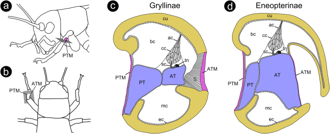
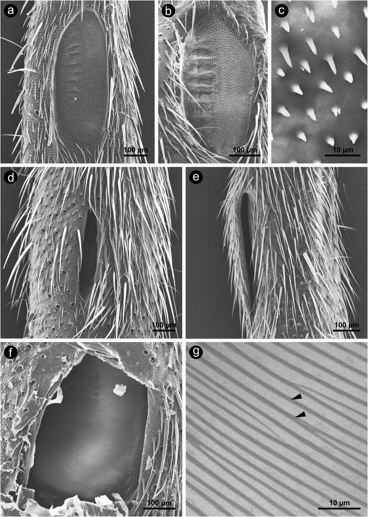
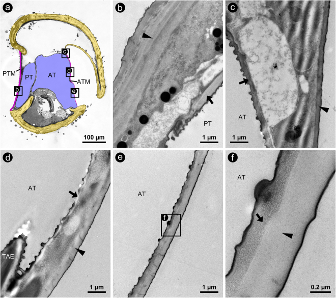
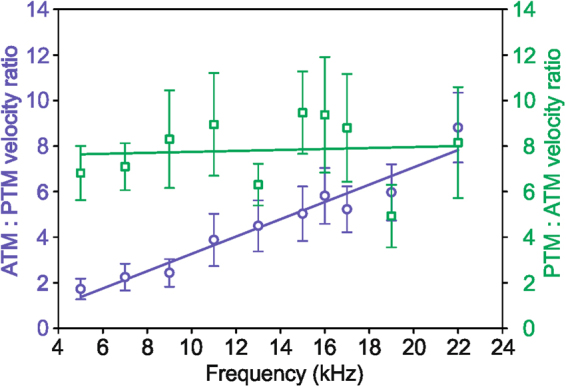
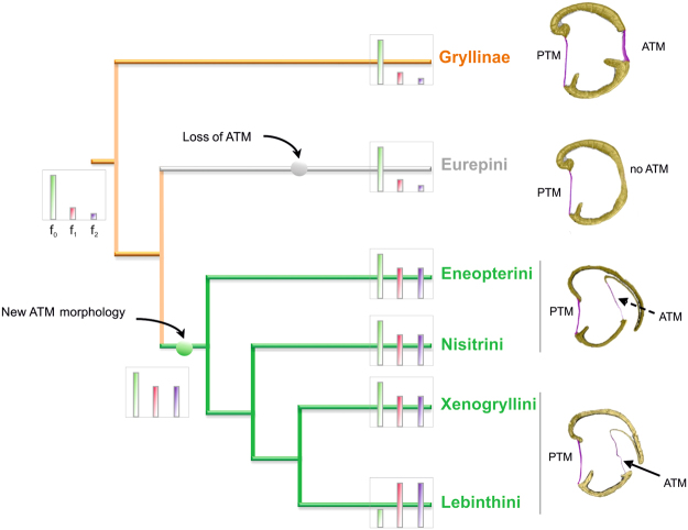
The receiver sensory system plays a crucial role in the evolution of new communication signals in insects. Among acoustic communicating crickets, the tribe Lebinthini (Eneopterinae) has evolved a unique communication system in that males produce exceptionally high-frequency calls and females respond with vibratory signals to guide males towards them. In this study, we describe nine species of Eneopterinae in which the sound receiving structures have undergone considerable morphological changes. We revealed that the anterior tympanal membrane (ATM) of the ear was extremely thin, as little as 0.35 µm thick, and to the best of our knowledge, this is the thinnest tympanal membrane found in crickets thus far. Measurements of tympanum vibrations obtained from Lebinthus bitaeniatus demonstrated a strong sensitivity towards higher frequencies. The finding also coincides with the neuronal tuning of ascending neurons and the behavioural response of the Lebinthini. The morphologically specialized ATM and its mechanical sensitivity for high frequencies, therefore, may have driven the sensory exploitation of an anti-predator behaviour that led to the evolution of a new communication system known for this group of crickets. The hypothetical phylogenetic origin of the investigated tympanal ears is discussed.


Conflict of interest statement

The authors declare that they have no competing interests.

Figures

Figure 1
Figure 1
Schematic drawings of tympanal hearing organs in crickets. (a) Cricket from side view and (b) from top view, illustrating the relative position and orientation of the cutting plane (grey rectangle) along which cross-sections through the hearing organs were made in the present study. (c) Schematic cross-section through a typical tympanal ear of the subfamily Gryllinae and (d) of the subfamily Eneopterinae, showing the main structural and sensory components. Orientation in (c and d): top = lateral; bottom = medial; left = posterior; right = anterior. Colour code: yellow = cuticular portion of the tibia; magenta = cuticular portion of the tympanal membranes; blue = interior of tracheal branches. Abbreviations: ac = attachment cell; AT = anterior tracheal branch; ATM = anterior tympanal membrane; bc = blood channel; cc = cap cell; cu = cuticle; ec = epidermal cell; mc = muscle channel; PT = posterior tracheal branch; PTM = posterior tympanal membrane; S = suspensorium; sc = sensory cell; tn = tympanal nerve.
Figure 2
Figure 2
SEM-micrographs of the proximal tibia of Eneopterinae crickets, showing different aspects of the posterior (PTM, ac) and anterior tympanum (ATM, dg). (a) PTM of G. baitabagus, (b) PTM of N. vittatus in overview and (c) at higher magnification, showing a uniform surface covered by microtrichia, (d) anterior view of the slitted ATM of M. jharnae, (e) medial view of the slitted ATM of N. vittatus, (f) anterior view of the ATM of C. sumba after removal of the cap that covers the ATM in the native state; overview (f) and at higher magnification (g), arrowheads indicate taenidia that are visible in SEM-micrographs because of the very low thickness of the ATM. Orientation in (ad,f and g): top = proximal; bottom = distal; left = lateral; right = medial. Orientation in (e): top = proximal; bottom = distal; left = anterior; right = posterior.
Figure 3
Figure 3
Coloured LM-micrographs of semi-thin cross-sections through the proximal tibia of (a) G. bimaculatus (Gryllinae) and of several cricket species of different tribes of the subfamily Eneopterinae (be). (b) E. guyanensis (tribe Eneopterini); (c) N. vittatus (tribe Nisitrini); (d) X. marmoratus (tribe Xenogryllini); (e) L. bitaeniatus (tribe Lebinthini). Note that for E. guyanensis (b) and X. marmoratus (d) only ethanol-fixed and air dried material was available, respectively. Tissue preservation in these species, therefore, was very poor. Thus, absolute and relative proportions of the internal compartments of the ears shown in (b) and (d) may not reflect the situation in the living animals (see e.g. the strongly shrunken muscle channel). Colour code: yellow = cuticular portion of the tibia; magenta = cuticular portion of the tympanal membranes; blue = interior of tracheal branches. Orientation: top = lateral; bottom = medial; left = posterior; right = anterior. Abbreviations: AT = anterior tracheal branch; ATM = anterior tympanal membrane; PT = posterior tracheal branch; PTM = posterior tympanal membrane.
Figure 4
Figure 4
Micrographs of cross-sections through the proximal tibia of L. bitaeniatus. (a) LM-micrograph of a semithin cross-section gives an overview on the position and orientation of the following subfigures indicated by lettered rectangles. TEM-micrographs of the PTM (b) and of the ATM at different locations (c–f); positions are indicated in subfigure a. Arrows and arrowheads indicate cuticular portions of the tracheal branches and of the tympanal membranes, respectively. Orientation: top = lateral; bottom = medial; left = posterior; right = anterior. Abbreviations: AT = anterior tracheal branch; ATM = anterior tympanal membrane; PT = posterior tracheal branch; PTM = posterior tympanal membrane; TAE = taenidium.
Figure 5
Figure 5
For the field cricket G. bimaculatus and the Eneopterinae species L. bitaeniatus, Laser Doppler vibrometry was used to determine the vibration velocities of the anterior (a and c) and posterior tympanum (b and d). Individual sensitivity tuning curves are shown as thin black lines and the respective mean curves (± standard error of mean) are shown as coloured, thick lines. The power spectrum (given in relative amplitude) of a typical male’s call are shown in the background of the subfigures (b) and (c).
Figure 6
Figure 6
Tympanal membrane velocity ratios were determined for G. bimaculatus (green squares, N = 7) and L. bitaeniatus (blue circles, N = 10); data are means ± standard error of means. A linear regression was fitted to the data, revealing highly significant frequency dependency for L. bitaeniatus (R2 = 0.939, p < 0.001, y = 0.38x) but not for G. bimaculatus (R2 = 0.006, p = 0.83, y = 0.02x). Note that the ratio of ATM:PTM velocity for L. bitaeniatus and the inverse ratio of PTM: ATM velocity for G. bimaculatus is shown.
Figure 7
Figure 7
Proposed phylogenetic origin of new ATM morphology. Phylogenetic tree showing the position of the subfamily Gryllinae and tribes of the subfamily Eneopterinae (derived from), including the representative ATM morphology and the characteristic spectrum of the calling song with the relative amount of acoustic energy of the dominant frequency (f0) and of the first two harmonics (f1 and f2). In the Lebinthini, the dominant frequency shifted either to f1 or f2. Compared to the Xenogryllini and Lebinthini, the Eneopterini and Nisitrini possess a cuticular cap that completely covered the chamber above the ATM. Representatives of the Eurepini were not included in this study, since they do not possess an ATM.

Similar articles

Cited by

References

    1. Hoy RR, Robert D. Tympanal hearing in insects. Annual Review of Entomology. 1996;41:433–450. doi: 10.1146/annurev.en.41.010196.002245. - DOI - PubMed
    1. Montealegre-Z. F, Jonsson T, Robson-Brown KA, Postles M, Robert D. Convergent evolution between insect and mammalian audition. Science. 2012;338:968–971. doi: 10.1126/science.1225271. - DOI - PubMed
    1. Yager DD. Structure, development, and evolution of insect auditory systems. Microscopy research and Technique. 1999;47:380–400. doi: 10.1002/(SICI)1097-0029(19991215)47:6<380::AID-JEMT3>3.0.CO;2-P. - DOI - PubMed
    1. Greenfield, M. D. Evolution of acoustic communication in insects in InsectHearing (eds. Pollack, G. S. et al.) 17–47 (Springer, 2016).
    1. Fullard JH, Yack JE. The evolutionary biology of insect hearing. Trends in Ecology & Evolution. 1993;8:248–252. doi: 10.1016/0169-5347(93)90200-9. - DOI - PubMed

Publication types