

Corneal Endothelial Cell Loss 3 Years After Successful Descemet Stripping Automated Endothelial Keratoplasty in the Cornea Preservation Time Study: A Randomized Clinical Trial
- PMID: 29127432
- PMCID: PMC6583548
- DOI: 10.1001/jamaophthalmol.2017.4970
Corneal Endothelial Cell Loss 3 Years After Successful Descemet Stripping Automated Endothelial Keratoplasty in the Cornea Preservation Time Study: A Randomized Clinical Trial
Abstract
Importance: Demonstrating that endothelial cell loss following Descemet stripping automated endothelial keratoplasty (DSAEK) is independent of donor cornea preservation time (PT) could increase the pool of corneal tissue available for keratoplasty.
Objective: To determine whether endothelial cell loss 3 years after successful DSAEK is related to PT.
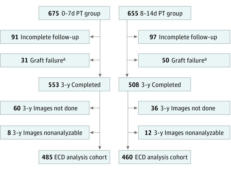
Design, setting, and participants: A multicenter, double-masked, randomized clinical trial included 40 clinical sites (70 surgeons) in the United States, with donor corneas provided by 23 US eye banks. A total of 945 eyes of 769 participants were included in the Cornea Preservation Time Study that had not experienced graft failure 3 years after DSAEK, performed primarily for Fuchs endothelial corneal dystrophy (96% of the cohort). The study was conducted from April 16, 2012, to June 5, 2017.
Interventions: DSAEK with random assignment of a donor cornea with PT of 0 to 7 days (0-7d PT) or 8 to 14 days (8-14d PT).
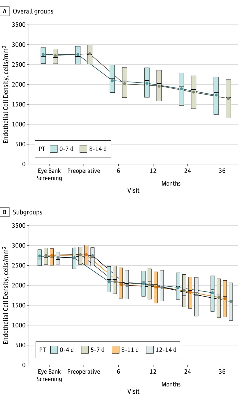
Main outcomes and measures: Endothelial cell density (ECD) at 3 years determined by a central image analysis reading center from clinical specular or confocal central endothelial images.
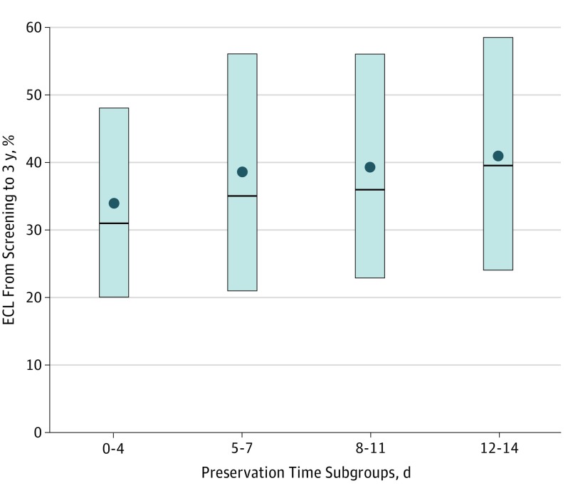
Results: Nine hundred forty-five eyes of 769 participants (median age, 70 years [range, 42-90 years], 60.8% women, 93.0% white) in the Cornea Preservation Time Study that had not experienced graft failure 3 years after DSAEK were included. At the initial eye bank tissue screening, mean (SD) central ECD was 2746 (297) cells/mm2 in the 0-7d PT group (n = 485) and 2723 (284) cells/mm2 in the 8-14d PT group (n = 460). At 3 years, the mean (SD) ECD decreased from baseline by 37% (21%) in the 0-7d PT group and 40% (22%) in the 8-14d PT group to 1722 (626) cells/mm2 and 1642 (631) cells/mm2, respectively (mean difference, 73 cells/mm2; 95% CI, 8-138 cells/mm2; P = .03). When analyzed as a continuous variable (days), longer PT was associated with lower ECD (mean difference by days, 15 cells/mm2; 95% CI, 4-26 cells/mm2; P = .006). Endothelial cell loss (ECL) was comparable from 4 to 13 days’ PT (n = 878; 36%-43% when tabulated by day). Available extension study ECD results at 4 years mirrored those at 3 years in the 203 eyes in the 0-7d PT group (mean [SD] ECD, 1620 [673] cells/mm2 and mean [SD] ECL, 41% [23%]) and 209 eyes in the 8-14d PT group (mean [SD] ECD, 1537 [683] cells/mm2 and mean [SD] ECL, 44% [23%]) (mean difference, 112 cells/mm2; 95% CI, 5-219 cells/mm2; P = .04).
Conclusions and relevance: Although ECL 3 years after Descemet stripping automated endothelial keratoplasty is greater with longer PT, the effect of PT on ECL is comparable from 4 to 13 days’ PT.
Trial registration: ClinicalTrials.gov NCT01537393.
Conflict of interest statement
Figures
References
-
- Waring GO III, Bourne WM, Edelhauser HF, Kenyon KR. The corneal endothelium: normal and pathologic structure and function. Ophthalmology. 1982;89(6):531-590. - PubMed
-
- Edelhauser HF. The resiliency of the corneal endothelium to refractive and intraocular surgery. Cornea. 2000;19(3):263-273. - PubMed
-
- Laule A, Cable MK, Hoffman CE, Hanna C. Endothelial cell population changes of human cornea during life. Arch Ophthalmol. 1978;96(11):2031-2035. - PubMed
-
- Zhang J, Patel DV. The pathophysiology of Fuchs’ endothelial dystrophy—a review of molecular and cellular insights. Exp Eye Res. 2015;130(1):97-105. - PubMed
-
- Park J, Yum HR, Kim MS, Harrison AR, Kim EC. Comparison of phaco-chop, divide-and-conquer, and stop-and-chop phaco techniques in microincision coaxial cataract surgery. J Cataract Refract Surg. 2013;39(10):1463-1469. - PubMed
Publication types
MeSH terms
Associated data
LinkOut - more resources
Full Text Sources
Other Literature Sources
Medical
Miscellaneous
