Review
. 2018 Apr:184:98-111.
doi: 10.1016/j.pharmthera.2017.11.002. Epub 2017 Nov 8.

Readers of DNA methylation, the MBD family as potential therapeutic targets

Affiliations
Review

Readers of DNA methylation, the MBD family as potential therapeutic targets

Gordon D Ginder et al. Pharmacol Ther. 2018 Apr.

Erratum in

Abstract

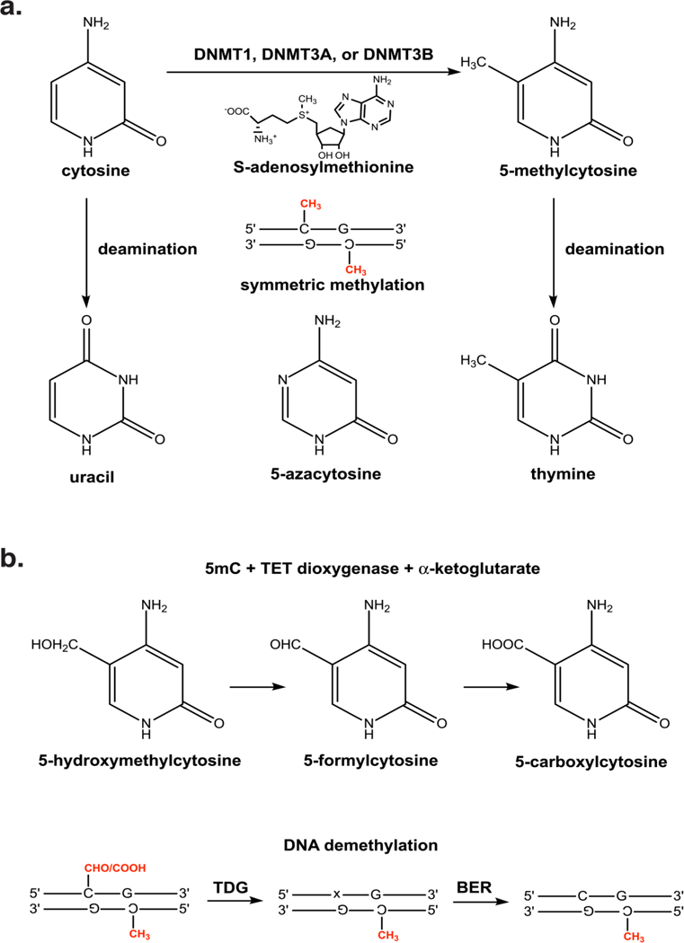
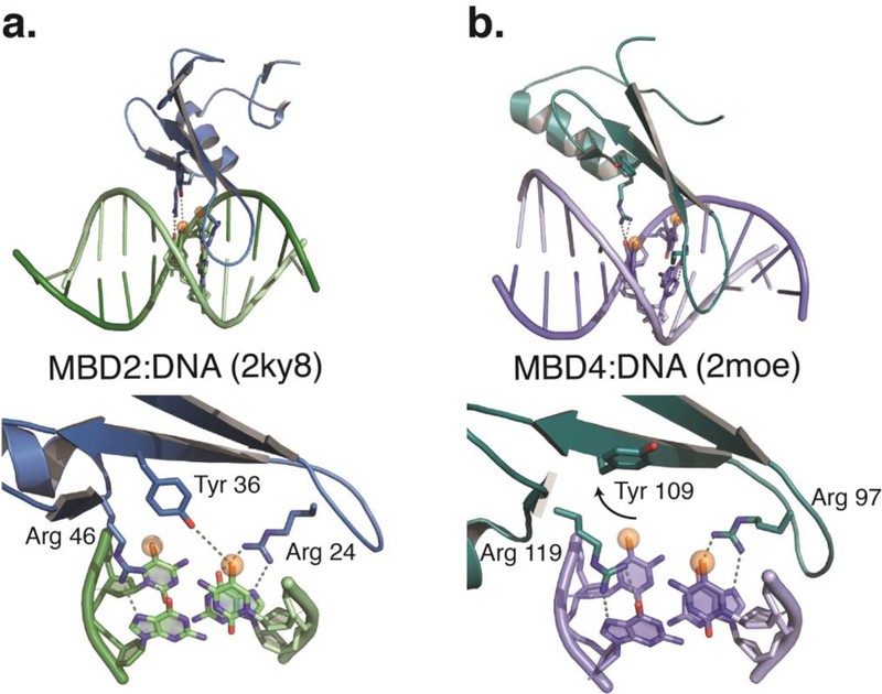
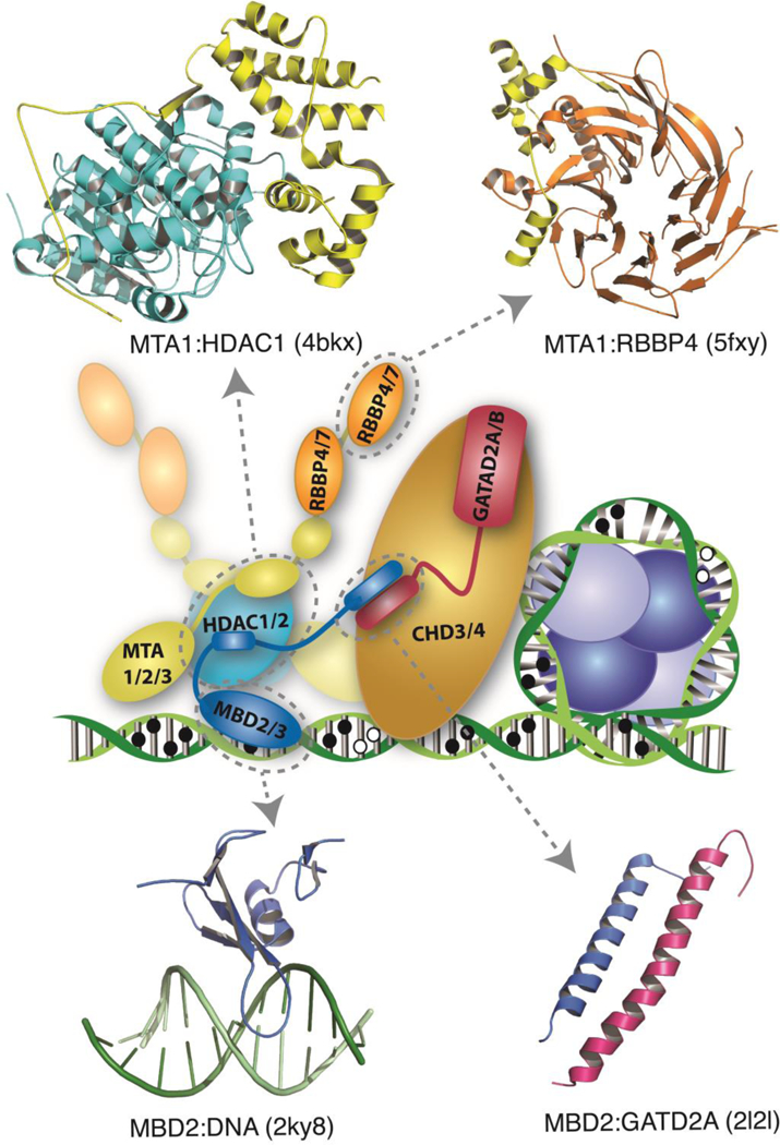
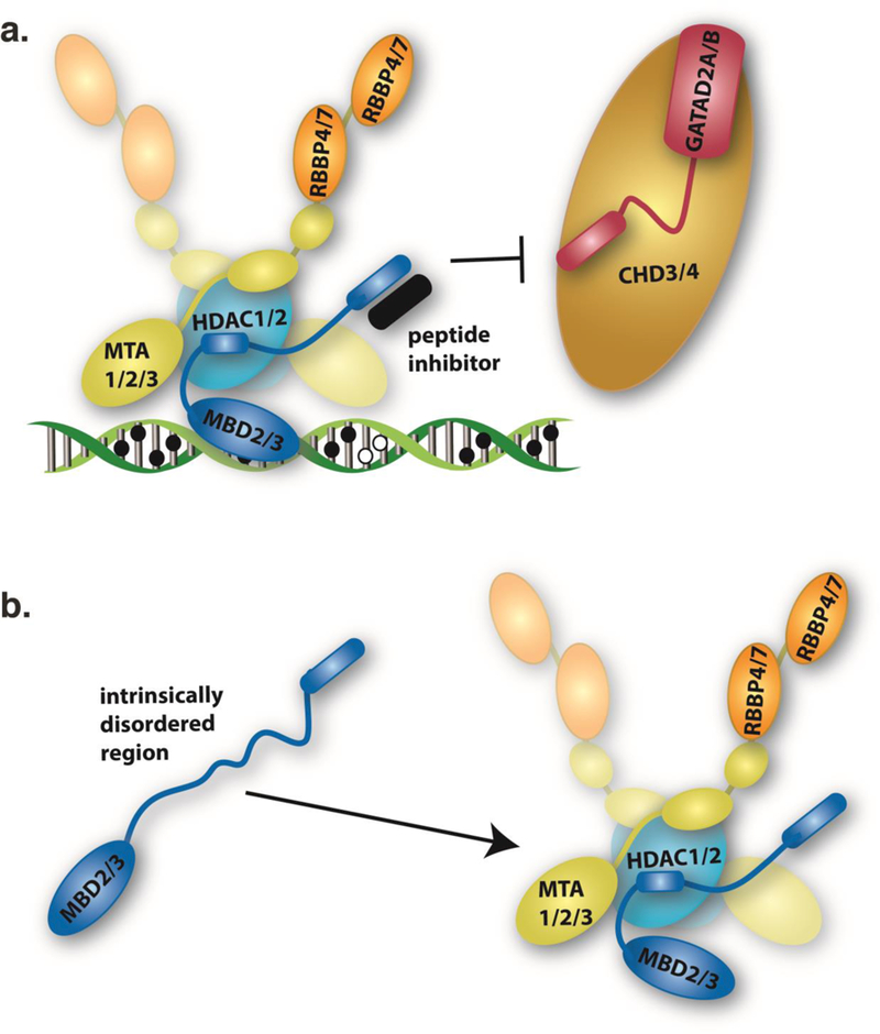
DNA methylation represents a fundamental epigenetic modification that regulates chromatin architecture and gene transcription. Many diseases, including cancer, show aberrant methylation patterns that contribute to the disease phenotype. DNA methylation inhibitors have been used to block methylation dependent gene silencing to treat hematopoietic neoplasms and to restore expression of developmentally silenced genes. However, these inhibitors disrupt methylation globally and show significant off-target toxicities. As an alternative approach, we have been studying readers of DNA methylation, the 5-methylcytosine binding domain family of proteins, as potential therapeutic targets to restore expression of aberrantly and developmentally methylated and silenced genes. In this review, we discuss the role of DNA methylation in gene regulation and cancer development, the structure and function of the 5-methylcytosine binding domain family of proteins, and the possibility of targeting the complexes these proteins form to treat human disease.

Keywords: 5-Methylcytosine binding domain; Chromatin; DNA methylation; Gene regulation; NuRD.


Conflict of interest statement

Conflict of Interest Statement: The authors declare that there are no conflicts of interest.

Figures

Fig. 1.
Fig. 1.
a) The DNMT enzymes add a methyl group to carbon-5 of symmetrically related cytosine bases in a CpG. Spontaneous deamination of cytosine and 5-methylcytosine generate uracil and thymine, respectively. Hence, deamination of 5-methylcytosine is a common cause of C → T and G → A transition mutations. 5-azacytosine contains a nitrogen at position 5 of the base which, when incorporated into DNA, inhibits methylation by the DNMT enzymes, b) The TET dioxygenase enzymes oxidize the methyl group of 5-methylcytosine to generate 5-hydroxymethylcytosine, 5- formylcytosine, and 5-carboxylcytosine. The latter two oxidation products can be excised by TDG, possibly in conjunction with other components of the base excision repair (BER) pathway, to form an abasic site (x) and demethylate the DNA.
Fig. 2.
Fig. 2.
a) A cartoon diagram depicts the solution structure of the MBD from MBD2 bound to methylated DNA (Scarsdale, et al., 2011). The domain contains a 3–4 strand β-sheet with a long finger like projection that extends down the major groove and a single α-helix. An expanded view shows how a tyrosine and two arginine residues from critical interactions with the 5-methylcytosine and two guanosine bases, respectively, of the mCpG. b) For comparison, cartoon diagram of the solution structure of MBD4 bound to methylated DNA (Walavalkar, et al., 2014) shows that the α-helix contains one additional turn, but otherwise has a similar structure. However, an expanded view reveals that the critical tyrosine residue rotates away from the DNA and does not interact with 5-methylcytosine. This structural rearrangement likely contributes to reduced selectivity for methylated DNA.
Fig. 3.
Fig. 3.
A schematic diagram shows the NuRD complex bound to methylated DNA next to a nucleosome. The complex comprises at least six core proteins, each of which have multiple paralogues (MBD2 or 3, in blue; MTA1,2, or 3, in yellow; HDAC1 or 2 in cyan; RBBP4 or 7, in orange; GATAD2A or B, in red; CHD3 or 4, in gold). While the structure of the full complex has not been determined to date, structures of several key interactions have been reported. Cartoon diagrams, generated with PyMOL (Schrodinger), are shown for structures that have been solved of the MTA1:HDAC1 (PDB ID - 4bkx) (Millard, et al., 2013), MTA1:RBBP4 (PDB ID - 5fxy) (Millard, et al., 2016), MBD2:DNA (PDB ID - 2ky8) (Scarsdale, et al., 2011), and MBD2:GATAD2A (PDB ID - 2l2l) (Gnanapragasam, et al., 2011) sub-complexes. Both stoichiometric (Acuna-Hidalgo, et al., 2017) and structural analyses indicate that the MTA1/2/3:HDAC1/2 sub-complex forms a dimer (Millard, et al., 2013), at least two RBBP4/7 bind to each MTA1/2/3 (Alqarni, et al., 2014; Schmidberger, et al., 2016), one to two copies of MBD2/3 and GATAD2A/B, and only copy of CHD3/4 are present in the complex.
Fig. 4.
Fig. 4.
a) A schematic cartoon depicts a peptide inhibitor (black) of complex NuRD complex formation. We previously demonstrated that a coiled-coil peptide, derived from the GATAD2A coiled-coil domain, restores expression of fetal/embryonic hemoglobin in tissue culture models of globin regulation (Gnanapragasam, et al., 2011). Immunoprecipitation of this peptide indicates that it disrupts function by blocking recruitment of CHD3/4 to the complex. b) Recently, we found that an intrinsically disordered region (IDR) of MBD2 is necessary and sufficient to bind the histone deacetylase core complex of NuRD comprising MTA1/2/3, HDAC1/2, and RBBP4/7 (Desai, et al., 2015). Secondary structure propensity of the isolated IDR suggests that it forms a helix upon interacting with the complex. Mutation of two consecutive residues within the helical region of the IDR disrupts interaction with the NuRD complex and abrogates methylation dependent silencing by MBD2-NuRD. Importantly, IDR-protein interactions have proven amenable to small molecule inhibition (Vela & Marzo, 2015). Hence, the MBD2 IDR represents a potential target for small molecule inhibition of NuRD complex formation.

References

    1. Acuna-Hidalgo R, Sengul H, Steehouwer M, van de Vorst M, Vermeulen SH, Kiemeney L, Veltman JA, Gilissen C, & Hoischen A (2017). Ultra-sensitive Sequencing Identifies High Prevalence of Clonal Hematopoiesis-Associated Mutations throughout Adult Life. Am J Hum Genet, 101, 50–64. - PMC - PubMed
    1. Aguilera C, Nakagawa K, Sancho R, Chakraborty A, Hendrich B, & Behrens A (2011). c-Jun N-terminal phosphorylation antagonises recruitment of the Mbd3/NuRD repressor complex. Nature, 469, 231–235. - PubMed
    1. Alqarni SS, Murthy A, Zhang W, Przewloka MR, Silva AP, Watson AA, Lejon S, Pei XY, Smits AH, Kloet SL, Wang H, Shepherd NE, Stokes PH, Blobel GA, Vermeulen M, Glover DM, Mackay JP, & Laue ED (2014). Insight into the architecture of the NuRD complex: structure of the RbAp48-MTA1 subcomplex. J Biol Chem, 289, 21844–21855. - PMC - PubMed
    1. Amaya M, Desai M, Gnanapragasam MN, Wang SZ, Zu Zhu S, Williams DC Jr., & Ginder GD (2013). Mi2beta-mediated silencing of the fetal gamma-globin gene in adult erythroid cells. Blood, 121, 3493–3501. - PMC - PubMed
    1. Amir RE, Van den Veyver IB, Wan M, Tran CQ, Francke U, & Zoghbi HY (1999). Rett syndrome is caused by mutations in X-linked MECP2, encoding methyl-CpG-binding protein 2. Nat Genet, 23, 185–188. - PubMed

Publication types

Substances