. 2018 Jan:65:216-225.
doi: 10.1016/j.actbio.2017.11.015. Epub 2017 Nov 8.

The hierarchical response of human corneal collagen to load

Affiliations

The hierarchical response of human corneal collagen to load

J S Bell et al. Acta Biomater. 2018 Jan.

Abstract

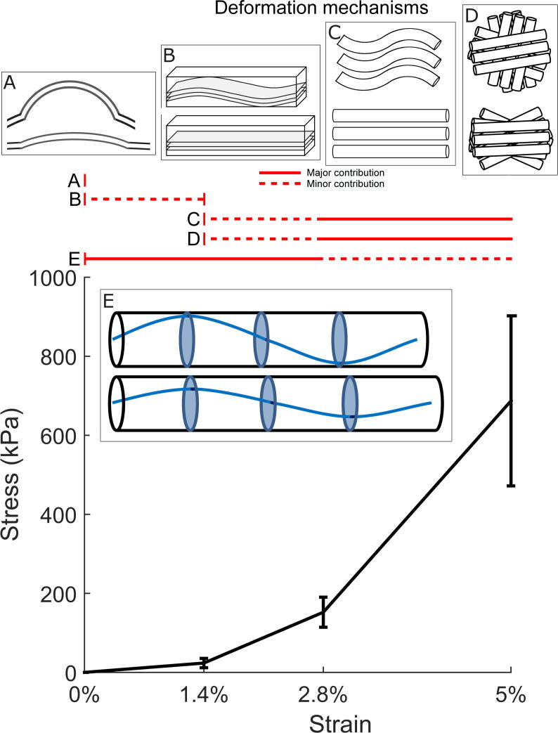
Fibrillar collagen in the human cornea is integral to its function as a transparent lens of precise curvature, and its arrangement is now well-characterised in the literature. While there has been considerable effort to incorporate fibrillar architecture into mechanical models of the cornea, the mechanical response of corneal collagen to small applied loads is not well understood. In this study the fibrillar and molecular response to tensile load was quantified using small and wide angle X-ray scattering (SAXS/WAXS), and digital image correlation (DIC) photography was used to calculate the local strain field that gave rise to the hierarchical changes. A molecular scattering model was used to calculate the tropocollagen tilt relative to the fibril axis and changes associated with applied strain. Changes were measured in the D-period, molecular tilt and the orientation and spacing of the fibrillar and molecular networks. These measurements were summarised into hierarchical deformation mechanisms, which were found to contribute at varying strains. The change in molecular tilt is indicative of a sub-fibrillar "spring-like" deformation mechanism, which was found to account for most of the applied strain under physiological and near-physiological loads. This deformation mechanism may play an important functional role in tissues rich in fibrils of high helical tilt, such as skin and cartilage.

Statement of significance: Collagen is the primary mediator of soft tissue biomechanics, and variations in its hierarchical structure convey the varying amounts of structural support necessary for organs to function normally. Here we have examined the structural response of corneal collagen to tensile load using X-rays to probe hierarchies ranging from molecular to fibrillar. We found a previously unreported deformation mechanism whereby molecules, which are helically arranged relative to the axis of their fibril, change in tilt akin to the manner in which a spring stretches. This "spring-like" mechanism accounts for a significant portion of the applied deformation at low strains (<3%). These findings will inform the future design of collagen-based artificial corneas being developed to address world-wide shortages of corneal donor tissue.

Keywords: Biomechanics; Collagen; Cornea; Microstructure; X-ray scattering.


Figures

None
Graphical abstract
Fig. 1
Fig. 1
Hierarchical arrangement of collagen in the cornea. Subfibrillar illustrations are not to scale – see for an atomistic interpretation. Abbreviations: MF – microfibril; TC – tropocollagen molecule. (Transmission electron microscopy image courtesy of Nada Aldahlawi, unpublished work).
Fig. 2
Fig. 2
Illustrations of X-ray diffraction images and peak analysis. A. Example SAXS scatter pattern. B. Example WAXS scatter pattern. C Radial SAXS plot corresponding to panel A. D. Radial WAXS plot corresponding to panel B. Labelled peaks: Interfibrillar spacing (IF); Meridionals 3 and 5 (M3, M5); Bessel-function shaped fibril cylinder transform (used to measure fibril diameter) (B); Intermolecular spacing (IM). Dotted and dashed lines correspond to exponential background subtractions.
Fig. 3
Fig. 3
Illustrations of the experiment and analysis protocols. A. Schematic of the extensometry experiment with scan positions overlaid on the strip. B. Example SAXS pattern showing regions defined as parallel and perpendicular to the applied strain, for axial scattering (the regions are swapped for lateral scattering). C. Corresponding radial intensity plot for B, showing meridional peaks 3–8 as an example. D Example WAXS pattern with overlaid calculation for polar dependence of intermolecular peak. E. The azimuthal distribution of signal in the intermolecular peak in panel D reveals the polar distribution of molecules (after reflection-based removal of gridline). The process is the same for calculating the fibril distribution from SAXS images. Insert: Polar patch plot of the molecular distribution shown in panel E, used for compactness when showing multiple distributions across a specimen.
Fig. 4
Fig. 4
Averaged change in X-ray scattering metrics associated with strain. Error bars show SEM and are offset in x to avoid overlap. Parallel and perpendicular fibril diameters could not be ascertained due to loss of peak shape when strained.
Fig. 5
Fig. 5
Representative polar patch plots of collagen fibril orientation calculated from the interfibrillar peak distribution observed using SAXS (A) and the intermolecular peak distribution observed using WAXS (B), for representative specimens at varying strain. The anisotropy value is the ratio of maximum parallel collagen to maximum orthogonal collagen. Some SAXS plots are noisy or omitted due to loss in signal associated with disorder at the fibrillar hierarchy.
Fig. 6
Fig. 6
Distribution of local strain and associated reorientation for a representative tensile strip. x-axes of plots coincide. A. Original image of the tensile strip at 5% applied strain with local longitudinal strain field obtained using DIC overlaid. The strain field data are binned into pixels 300 µm in size. B. Average parallel and perpendicular strain along the central longitudinal/parallel axis of the strip, calculated from the central two rows of DIC pixels. C. Polar patch plots of local collagen molecule orientation at 5% applied strain. D. Anisotropy measured as ratio of peak longitudinal to peak orthogonal orientation, for each strain increment.
Fig. 7
Fig. 7
Molecular model geometry and results. A. Axial view of representative line/filament arrangement, which is a disordered hexagonal lattice. B. Illustration of how a helical molecular arrangement (blue) relative to the fibril axis (black) is modelled by a sum of straight line geometries of varying inclination. C. Side view of line model geometry, with molecular tilt shown as inclination angle θ. For each value of θ, 180 scatter patterns were calculated at 2° azimuthal increments to simulate an arrangement of parallel helices. D. Scatter pattern from the disordered helix model with a tilt of 16°. E. Modelled scatter pattern from a helical model with tilt 16°, convoluted with the average fibril distribution at the tare load. F. Representative WAXS pattern from a specimen under the tare load. G. Averaged polar arrangement of fibrils (blue), molecules (red) and modelled molecules at a 16° tilt (black) for a specimen under the tare load. F. Polar arrangements for a specimen at 5% applied strain, with a fitted 11° tilt.
Fig. 8
Fig. 8
Average quasi-equilibrium tensile stress associated with each strain increment, with spans indicating ranges of strain over which illustrated deformation mechanisms may apply. A. Straightening of corneal curvature, occurs during application of tare load (millimetres). B. Lamellar crimp, arguable contribution at low strains based upon literature evidence , (100 s of microns). C. Fibrillar crimp measured using SAXS (100 s of nanometres). D. Lamellar and fibrillar reorientation measured using SAXS (100 s of nanometres). E. Straightening of molecular helical tilt relative to fibril axis measured using SAXS and WAXS (Angstroms).

References

    1. Sasaki N., Odajima S. Stress-strain curve and Young’s modulus of a collagen molecule as determined by the X-ray diffraction technique. J. Biomech. 1996;29:655–658. - PubMed
    1. Bruckner P. Suprastructures of extracellular matrices: paradigms of functions controlled by aggregates rather than molecules. Cell Tissue Res. 2010;339:7–18. - PubMed
    1. Kadler K.E., Baldock C., Bella J., Boot-Handford R.P. Collagens at a glance. J. Cell Sci. 2007;120:1955–1958. - PubMed
    1. Orgel J.P.R.O., Irving T.C., Miller A., Wess T.J. Microfibrillar structure of type I collagen in situ. Proc. Natl. Acad. Sci. 2006;103:9001–9005. - PMC - PubMed
    1. Holmes D.F., Gilpin C.J., Baldock C., Ziese U., Koster A.J., Kadler K.E. Corneal collagen fibril structure in three dimensions: structural insights into fibril assembly, mechanical properties, and tissue organization. Proc. Natl. Acad. Sci. 2001;98:7307–7312. - PMC - PubMed

Substances

LinkOut - more resources