

Quantification and phenotypic characterisation of peripheral IFN-γ producing leucocytes in chickens vaccinated against Newcastle disease
- PMID: 29129224
- PMCID: PMC5697524
- DOI: 10.1016/j.vetimm.2017.10.001
Quantification and phenotypic characterisation of peripheral IFN-γ producing leucocytes in chickens vaccinated against Newcastle disease
Abstract
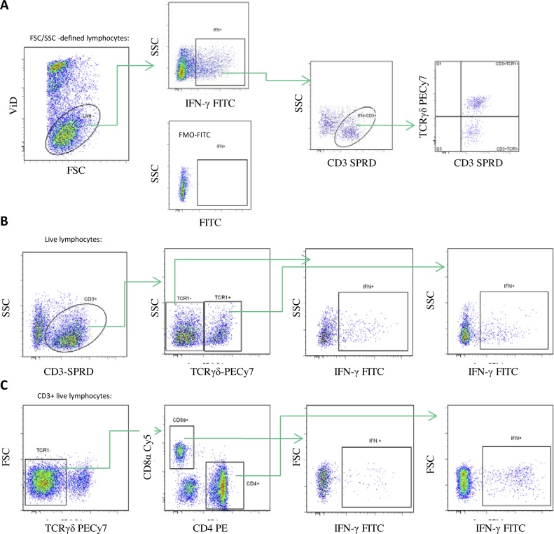
The aim of this study was to optimise and evaluate an intracellular cytokine staining (ICS) assay for assessment of T cell IFN-γ responses in chickens vaccinated against Newcastle disease (ND). We aimed to validate currently available antibodies to chicken IFN-γ using transfected CHO cells. Moreover, this ICS assay was evaluated for use to detect mitogen and antigen induced IFN-γ production in chicken peripheral blood leucocytes. Chickens from an inbred white leghorn line containing two MHC haplotypes, B19 and B21, were divided into three experimental groups; one group was kept as naive controls, one group was vaccinated intramuscularly twice with a commercial inactivated ND virus (NDV) vaccine, and the last group was vaccinated orally twice with a commercial live attenuated NDV vaccine. PBMC were ex vivo stimulated with ConA or with NDV antigen. The ICS assay was used to determine the phenotype and frequency of IFN-γ positive cells. ConA stimulation induced extensive IFN-γ production in both CD3+TCRγδ+ (γδ T cells) cells and CD3+TCRγδ- cells (αβ T cells), but no significant differences were observed between the experimental groups. Furthermore, a large proportion of the IFN-γ producing cells were CD3- indicating that other cells than classic T cells, secreted this cytokine. NDV antigen stimulation induced IFN-γ production but to a lower extent than ConA and with a large variation between individuals. The CD3+TCR1γδ-CD8α+ (CTL) population produced the highest NDV specific IFN-γ responses, with significantly elevated levels of IFN-γ producing cells in the B19 chickens vaccinated orally with live attenuated NDV vaccine. This was not the case in the B21 animals, indicating a haplotype restricted variation. In contrast, the CD3+TCR1γδ-CD4+ (Th) population did not show a significant increase in IFN-γ production in NDV stimulated samples which was in part due to a high number of IFN-γ producing cells after incubation with medium alone. In conclusion, an ICS assay for phenotyping of IFN-γ producing chicken leukocytes was set up that proved useful in identifying cytokine producing cells upon either mitogen or antigen-specific stimulation.
Keywords: Chicken; Flow cytometry; Interferon-γ; Intracellular cytokine staining; T cells; Vaccines.
Copyright © 2017 The Authors. Published by Elsevier B.V. All rights reserved.
Figures


Similar articles
-
Immunomodulation of bivalent Newcastle disease DNA vaccine induced immune response by co-delivery of chicken IFN-γ and IL-4 genes.Vet Immunol Immunopathol. 2011 Nov 15;144(1-2):36-44. doi: 10.1016/j.vetimm.2011.07.006. Epub 2011 Jul 19. Vet Immunol Immunopathol. 2011. PMID: 21820185
-
Assessment of the cell-mediated immune response in chickens by detection of chicken interferon-gamma in response to mitogen and recall Newcastle disease viral antigen stimulation.Avian Pathol. 2004 Jun;33(3):343-50. doi: 10.1080/0307945042000220318. Avian Pathol. 2004. PMID: 15223565
-
Expression of interferon gamma by a highly virulent strain of Newcastle disease virus decreases its pathogenicity in chickens.Microb Pathog. 2013 Aug-Sep;61-62:73-83. doi: 10.1016/j.micpath.2013.05.009. Epub 2013 May 25. Microb Pathog. 2013. PMID: 23711962
-
Flow cytometric assessment of antigen-specific proliferation in peripheral chicken T cells by CFSE dilution.Vet Immunol Immunopathol. 2010 Nov 15;138(1-2):85-94. doi: 10.1016/j.vetimm.2010.07.010. Epub 2010 Jul 22. Vet Immunol Immunopathol. 2010. PMID: 20739071
-
Flow cytometric assessment of chicken T cell-mediated immune responses after Newcastle disease virus vaccination and challenge.Vaccine. 2010 Jun 17;28(28):4506-14. doi: 10.1016/j.vaccine.2010.04.044. Epub 2010 Apr 29. Vaccine. 2010. PMID: 20434546
Cited by
-
Adjuvant Effects of Platycodin D on Immune Responses to Infectious Bronchitis Vaccine in Chickens.J Poult Sci. 2020 Apr 25;57(2):160-167. doi: 10.2141/jpsa.0180089. J Poult Sci. 2020. PMID: 32461731 Free PMC article.
-
Immune Response to Salmonella Enteritidis Infection in Broilers Immunized Orally With Chitosan-Based Salmonella Subunit Nanoparticle Vaccine.Front Immunol. 2020 May 19;11:935. doi: 10.3389/fimmu.2020.00935. eCollection 2020. Front Immunol. 2020. PMID: 32508828 Free PMC article.
-
Development of Molecular Mechanisms and Their Application on Oncolytic Newcastle Disease Virus in Cancer Therapy.Front Mol Biosci. 2022 Jul 4;9:889403. doi: 10.3389/fmolb.2022.889403. eCollection 2022. Front Mol Biosci. 2022. PMID: 35860357 Free PMC article. Review.
-
Flow Cytometric Evaluation of CD4+ and CD8+ T-cell Immune Response in SPF Chickens Induced by Fowlpox Vaccine.Arch Razi Inst. 2021 Sep 1;76(3):429-436. doi: 10.22092/ari.2020.343514.1508. eCollection 2021 Summer. Arch Razi Inst. 2021. PMID: 34824736 Free PMC article.
-
Cytokine production and phenotype of Histomonas meleagridis-specific T cells in the chicken.Vet Res. 2019 Dec 5;50(1):107. doi: 10.1186/s13567-019-0726-z. Vet Res. 2019. PMID: 31806018 Free PMC article.
References
-
- Alexander D.J. Gordon memorial lecture. Newcastle disease. Br Poult Sci . 2001;42:5–22. - PubMed
-
- Al-Garib S., Gielkens A., Gruys D., Hartog L., Koch G. Immunoglobulin class distribution of systemic and mucosal antibody responses to Newcastle disease in chickens. Avian Dis. 2003;47:32–40. - PubMed
-
- Ariaans M., van de Haar P., Lowenthal J., van E., Hensen E., Vervelde L. ELISPOT and intracellular cytokine staining: novel assays for quantifying T cell responses in the chicken. Dev. Comp. Immunol. 2008;32:1398–1404. - PubMed
-
- Breed D., Dorrestein J., Schetters T., Waart L., Rijke E., Vermeulen A. Peripheral blood lymphocytes from Eimeria tenella infected chickens produce gamma-interferon after stimulation in vitro. Parasite Immunol. 1997;19:127–135. - PubMed
Publication types
MeSH terms
Substances
Grants and funding
LinkOut - more resources
Full Text Sources
Other Literature Sources
Research Materials
