Glutamylation Regulates Transport, Specializes Function, and Sculpts the Structure of Cilia
- PMID: 29129530
- PMCID: PMC5698134
- DOI: 10.1016/j.cub.2017.09.066
Glutamylation Regulates Transport, Specializes Function, and Sculpts the Structure of Cilia
Abstract
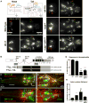
Ciliary microtubules (MTs) are extensively decorated with post-translational modifications (PTMs), such as glutamylation of tubulin tails. PTMs and tubulin isotype diversity act as a "tubulin code" that regulates cytoskeletal stability and the activity of MT-associated proteins such as kinesins. We previously showed that, in C. elegans cilia, the deglutamylase CCPP-1 affects ciliary ultrastructure, localization of the TRP channel PKD-2 and the kinesin-3 KLP-6, and velocity of the kinesin-2 OSM-3/KIF17, whereas a cell-specific α-tubulin isotype regulates ciliary ultrastructure, intraflagellar transport, and ciliary functions of extracellular vesicle (EV)-releasing neurons. Here we examine the role of PTMs and the tubulin code in the ciliary specialization of EV-releasing neurons using genetics, fluorescence microscopy, kymography, electron microscopy, and sensory behavioral assays. Although the C. elegans genome encodes five tubulin tyrosine ligase-like (TTLL) glutamylases, only ttll-11 specifically regulates PKD-2 localization in EV-releasing neurons. In EV-releasing cephalic male (CEM) cilia, TTLL-11 and the deglutamylase CCPP-1 regulate remodeling of 9+0 MT doublets into 18 singlet MTs. Balanced TTLL-11 and CCPP-1 activity fine-tunes glutamylation to control the velocity of the kinesin-2 OSM-3/KIF17 and kinesin-3 KLP-6 without affecting the intraflagellar transport (IFT) kinesin-II. TTLL-11 is transported by ciliary motors. TTLL-11 and CCPP-1 are also required for the ciliary function of releasing bioactive EVs, and TTLL-11 is itself a novel EV cargo. Therefore, MT glutamylation, as part of the tubulin code, controls ciliary specialization, ciliary motor-based transport, and ciliary EV release in a living animal. We suggest that cell-specific control of MT glutamylation may be a conserved mechanism to specialize the form and function of cilia.
Keywords: C. elegans; cilia; extracellular vesicles; glutamylation; intraflagellar transport; kinesin-2; kinesin-3; microtubule; polycystin; post-translational modifications.
Copyright © 2017 Elsevier Ltd. All rights reserved.
Figures


Similar articles
-
The tubulin deglutamylase CCPP-1 regulates the function and stability of sensory cilia in C. elegans.Curr Biol. 2011 Oct 25;21(20):1685-94. doi: 10.1016/j.cub.2011.08.049. Epub 2011 Oct 6. Curr Biol. 2011. PMID: 21982591 Free PMC article.
-
Mutation of NEKL-4/NEK10 and TTLL genes suppress neuronal ciliary degeneration caused by loss of CCPP-1 deglutamylase function.PLoS Genet. 2020 Oct 16;16(10):e1009052. doi: 10.1371/journal.pgen.1009052. eCollection 2020 Oct. PLoS Genet. 2020. PMID: 33064774 Free PMC article.
-
Cell-Specific α-Tubulin Isotype Regulates Ciliary Microtubule Ultrastructure, Intraflagellar Transport, and Extracellular Vesicle Biology.Curr Biol. 2017 Apr 3;27(7):968-980. doi: 10.1016/j.cub.2017.02.039. Epub 2017 Mar 16. Curr Biol. 2017. PMID: 28318980 Free PMC article.
-
The tubulin code specializes neuronal cilia for extracellular vesicle release.Dev Neurobiol. 2021 Apr;81(3):231-252. doi: 10.1002/dneu.22787. Epub 2020 Nov 8. Dev Neurobiol. 2021. PMID: 33068333 Free PMC article. Review.
-
Mating behavior, male sensory cilia, and polycystins in Caenorhabditis elegans.Semin Cell Dev Biol. 2014 Sep;33:25-33. doi: 10.1016/j.semcdb.2014.06.001. Epub 2014 Jun 27. Semin Cell Dev Biol. 2014. PMID: 24977333 Free PMC article. Review.
Cited by
-
Tubulin code eraser CCP5 binds branch glutamates by substrate deformation.Nature. 2024 Jul;631(8022):905-912. doi: 10.1038/s41586-024-07699-0. Epub 2024 Jul 17. Nature. 2024. PMID: 39020174
-
CEP41-mediated ciliary tubulin glutamylation drives angiogenesis through AURKA-dependent deciliation.EMBO Rep. 2020 Feb 5;21(2):e48290. doi: 10.15252/embr.201948290. Epub 2019 Dec 29. EMBO Rep. 2020. PMID: 31885126 Free PMC article.
-
Impaired glutamylation of RPGRORF15 underlies the cone-dominated phenotype associated with truncating distal ORF15 variants.Proc Natl Acad Sci U S A. 2022 Dec 6;119(49):e2208707119. doi: 10.1073/pnas.2208707119. Epub 2022 Nov 29. Proc Natl Acad Sci U S A. 2022. PMID: 36445968 Free PMC article.
-
Cilia structure and intraflagellar transport differentially regulate sensory response dynamics within and between C. elegans chemosensory neurons.PLoS Biol. 2024 Nov 26;22(11):e3002892. doi: 10.1371/journal.pbio.3002892. eCollection 2024 Nov. PLoS Biol. 2024. PMID: 39591402 Free PMC article.
-
Regulation of phosphoribosyl ubiquitination by a calmodulin-dependent glutamylase.Nature. 2019 Aug;572(7769):387-391. doi: 10.1038/s41586-019-1439-1. Epub 2019 Jul 22. Nature. 2019. PMID: 31330531 Free PMC article.
References
-
- Fisch C, Dupuis-Williams P. Ultrastructure of cilia and flagella - back to the future! Biol Cell. 2011;103:249–270. - PubMed
-
- Konno A, Ikegami K, Konishi Y, Yang HJ, Abe M, Yamazaki M, Sakimura K, Yao I, Shiba K, Inaba K, et al. Ttll9−/− mice sperm flagella show shortening of doublet 7, reduction of doublet 5 polyglutamylation and a stall in beating. J Cell Sci. 2016;129:2757–2766. - PubMed
-
- Perkins LA, Hedgecock EM, Thomson JN, Culotti JG. Mutant sensory cilia in the nematode Caenorhabditis elegans. Dev Biol. 1986;117:456–487. - PubMed
-
- Tsuji T, Matsuo K, Nakahari T, Marunaka Y, Yokoyama T. Structural basis of the Inv compartment and ciliary abnormalities in Inv/nphp2 mutant mice. Cytoskeleton (Hoboken) 2016;73:45–56. - PubMed
MeSH terms
Substances
Grants and funding
LinkOut - more resources
Full Text Sources
Other Literature Sources
Research Materials
Miscellaneous

