. 2018 Feb;26(2):245-254.
doi: 10.1016/j.joca.2017.10.019. Epub 2017 Nov 9.

TSG-6 - a double-edged sword for osteoarthritis (OA)

Affiliations

TSG-6 - a double-edged sword for osteoarthritis (OA)

C-H Chou et al. Osteoarthritis Cartilage. 2018 Feb.

Abstract

Purpose: To explore mechanisms underlying the association of TSG-6 with osteoarthritis (OA) progression.

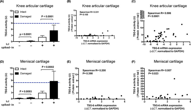
Methods: TSG-6-mediated heavy chain (HC) transfer (TSG-6 activity) and its association with inflammatory mediators were quantified in knee OA (n=25) synovial fluids (SFs). Paired intact and damaged cartilages from the same individuals (20 tibial and 12 meniscal) were analyzed by qRT-PCR and immunohistochemistry (IHC) for gene and protein expression of TSG-6 and components of Inter-alpha-Inhibitor (IαI) and TSG-6 activity ± spiked in IαI. Primary chondrocyte cultures (n=5) ± IL1β or TNFα were evaluated for gene expression. The effects of TSG-6 activity on cartilage extracellular matrix (ECM) assembly were explored using quantitative hyaluronan (HA)-aggrecan binding assays.

Results: TSG-6 activity was significantly associated (R > 0.683, P < 0.0002) with inflammatory mediators including TIMP-1, A2M, MMP3, VEGF, VCAM-1, ICAM-1 and IL-6. Although TSG-6 protein and mRNA were highly expressed in damaged articular and meniscal cartilage and cytokine-treated chondrocytes, there was little or no cartilage expression of components of the IαI complex (containing HC1). By IHC, TSG-6 was present throughout lesioned cartilage but HC1 only at lesioned surfaces. TSG-6 impaired HA-aggrecan assembly, but TSG-6 mediated HA-HC formation reduced this negative effect.

Conclusions: TSG-6 activity is a global inflammatory biomarker in knee OA SF. IαI, supplied from outside cartilage, only penetrates the cartilage surface, restricting TSG-6 activity (HC transfer) to this region. Therefore, unopposed TSG-6 in intermediate and deep regions of OA cartilage could possibly block matrix assembly, leading to futile synthesis and account for increased risk of OA progression.

Keywords: Biomarker; Cartilage matrix; Hyaluronan; Inflammation; Osteoarthritis progression; TSG-6.


Conflict of interest statement

Competing interest statement

The authors declare that they have no competing interests.

Figures

Figure 1
Figure 1. Correlations of TSG-6 activity and inflammatory mediators in human OA SF in EC20 cohort
(A–B) Correlation of TSG-6 activity with protease inhibitors: TIMP-1 (R=0.890, P<0.0001) A2M (R=0.831, P<0.0001). (C–E) Correlation of TSG-6 activity with inflammatory mediators: VEGF (R=0.734, and P<0.0001), VCAM-1 (R=0.730, and P<0.0001) and ICAM-1 (R=0.688, and P=0.0001). (F) Correlation of TSG-6 activity with MMP3 (protease). Spearman’s correlation R=0.763, and P<0.0001. (G) Correlation of TSG-6 activity with cytokine IL-6. Pearson’s correlation R=0.683, and P=0.0002). (H) TSG-6 activity in SFs significantly correlated with the concentration of TSG-6 protein as measured by ELISA (assessed using Spearman correlation, n=25). The markers that were fully measurable but that were not significantly associated with TSG-6 activity were listed in the supplementary table-1.
Figure 2
Figure 2. Expression of TSG-6 and IαI complex related genes in OA cartilage from joint replacement and primary chondrocytes with or without cytokines treatment
mRNAs were quantified by qRT-PCR in matched intact and damaged cartilages (20 tibial and 12 paired menisci) and primary chondrocytes (n=5) with or without IL1ß or TNFα treatment. TNFAIP6 (TSG-6) was highly and significantly differentially expressed in chondrocytes from damaged (red) compared to intact (blue) (A) tibial plateau cartilage and (B) meniscal cartilage and (C) primary chondrocytes treated with cytokines for 24 hrs. ITIH1, ITIH2, and AMBP (bikunin) were either not expressed or expressed at a low level in cartilage and chondrocytes, i.e. ~212 lower than GAPDH that was used as an endogenous control to normalize sample loading. NC represents the negative control (water in lieu of primers) with undetectable expression. FC=fold-change. P values were determined by paired t-test. Error bar = standard deviation.
Figure 3
Figure 3. Immunolocalization of HC1 and TSG-6 in HA-rich matrices of intact and damaged regions of OA cartilages including tibial plateau and meniscus
Antibodies against HC1 and TSG-6 were used to detect their protein expression (dark brown staining) by immunohistochemistry in the HA-rich matrices of cartilage from (A) tibial plateau and (B) meniscus. High expression levels of HC1 (yellow triangles) and TGS-6 (red arrows) were detected and co-localized on the surface of damaged cartilage, with little or none or much detected in intact cartilage. Non-immune IgGs were used as negative controls. All sections were counterstained light blue with hematoxylin. The scale bars are 0.3 mm.
Figure 4
Figure 4. Addition of exogenous IαI increases TSG-6 activity of cartilage
TSG-6 activities in protein extracts from cartilage with or without exogenous IαI and the correlations between TSG-6 activity and gene expression. Compared to intact cartilage, protein extracts from damaged cartilage had higher TSG-6 activities although the activities from both regions were low in (A) articular and (D) meniscal cartilage (intact articular 0.02±0.05 U, damaged articular 0.23±0.40 U; intact meniscal 0.20±0.35 U, damaged meniscal 0.46±0.57 U). With exogenous IαI spiked-in, TSG-6 activities of protein extracts increased significantly (p<0.0001 in each group between with and without IαI spiked-in) and were consistently higher in damaged compared with intact cartilage (intact articular 2.11±1.82 U, damaged articular 5.36±5.88 U; intact meniscal 2.79±1.80 U, damaged meniscal 8.57±9.51 U). Without IαI spiked-in, TSG-6 activity was not correlated with TSG-6 gene expression in the articular (B) or meniscal cartilage (E). With exogenous IαI spiked-in, TSG-6 activity increased and correlated with TSG-6 gene expression in both articular (C) and meniscal (F) cartilages. In addition, with exogenous IαI spiked-in, the TSG-6 activities of damaged cartilages were similar to the mean TSG-6 activity levels in OA synovial fluids (A and D, blue dotted lines). These data demonstrate that endogenous IαI is rate limiting for TSG-6 activity in cartilage. Error bar = standard deviation.
Figure 5
Figure 5
Formation of HA-HC results in retained ability of HA to bind HA binding proteins. The consequence of generating HA-HC (TSG-6 activity in the presence of IαI) for matrix assembly was examined using (A) HA binding protein (HABP) and (B) aggrecan (A1D1) binding assays. Pre-incubation of HA with increasing (A) or high (B) concentrations of TSG-6 significantly reduced binding of HABP and aggrecan to immobilized HA, respectively. HA-HC formation resulted in retained ability of immobilized HA to bind HABP and partially but significantly restored the ability of aggrecan to bind HA. Dotted and dashed lines in A represent the OD values of negative controls consisting of HA pre-incubated with buffer and 20nM IαI, respectively. These results suggest a negative effect of TSG-6 in the absence of IαI on matrix assembly capabilities. Paired t-test. **P<0.001; ***P<0.0001.
Figure-6
Figure-6. The potential role of TSG-6 in joint disease
TSG-6 can come from cartilage or synovial macrophages in response to inflammatory mediators, including IL-1ß and TNFα. The anti-plasmin effect of IαI-TSG-6 was minor due to the presence of HA in knee joints. High expression of TSG-6 in full thickness cartilage damaged by OA causes ECM disruption due to increasing HA condensation and reduced ECM assembly. In conjunction with IαI, TSG-6-mediated heavy chain transfer activity (TSG-6 activity) could mitigate the negative effects of TSG-6 on cartilage matrix assembly and turnover. TSG-6 may promote pathology in cartilage in OA knees. However, the surface disruption that occurs from OA damage exposes the cartilage to IαI from the synovial fluid and enables TSG-6 activity to mitigate the damage.

References

    1. Kehlen A, Pachnio A, Thiele K, Langner J. Gene expression induced by interleukin-17 in fibroblast-like synoviocytes of patients with rheumatoid arthritis: upregulation of hyaluronan-binding protein TSG-6. Arthritis Res Ther. 2003;5:R186–192. - PMC - PubMed
    1. Lee TH, Lee GW, Ziff EB, Vilcek J. Isolation and characterization of eight tumor necrosis factor-induced gene sequences from human fibroblasts. Mol Cell Biol. 1990;10:1982–1988. - PMC - PubMed
    1. Maier R, Wisniewski HG, Vilcek J, Lotz M. TSG-6 expression in human articular chondrocytes. Possible implications in joint inflammation and cartilage degradation. Arthritis Rheum. 1996;39:552–559. - PubMed
    1. Robinson LJ, Tourkova I, Wang Y, Sharrow AC, Landau MS, Yaroslavskiy BB, et al. FSH-receptor isoforms and FSH-dependent gene transcription in human monocytes and osteoclasts. Biochemical and biophysical research communications. 2010;394:12–17. - PMC - PubMed
    1. Yoshioka S, Ochsner S, Russell DL, Ujioka T, Fujii S, Richards JS, et al. Expression of tumor necrosis factor-stimulated gene-6 in the rat ovary in response to an ovulatory dose of gonadotropin. Endocrinology. 2000;141:4114–4119. - PubMed

Publication types