. 2018 Jan 18;25(1):78-87.e5.
doi: 10.1016/j.chembiol.2017.09.010. Epub 2017 Nov 9.

Lessons in PROTAC Design from Selective Degradation with a Promiscuous Warhead

Affiliations

Lessons in PROTAC Design from Selective Degradation with a Promiscuous Warhead

Daniel P Bondeson et al. Cell Chem Biol. .

Abstract

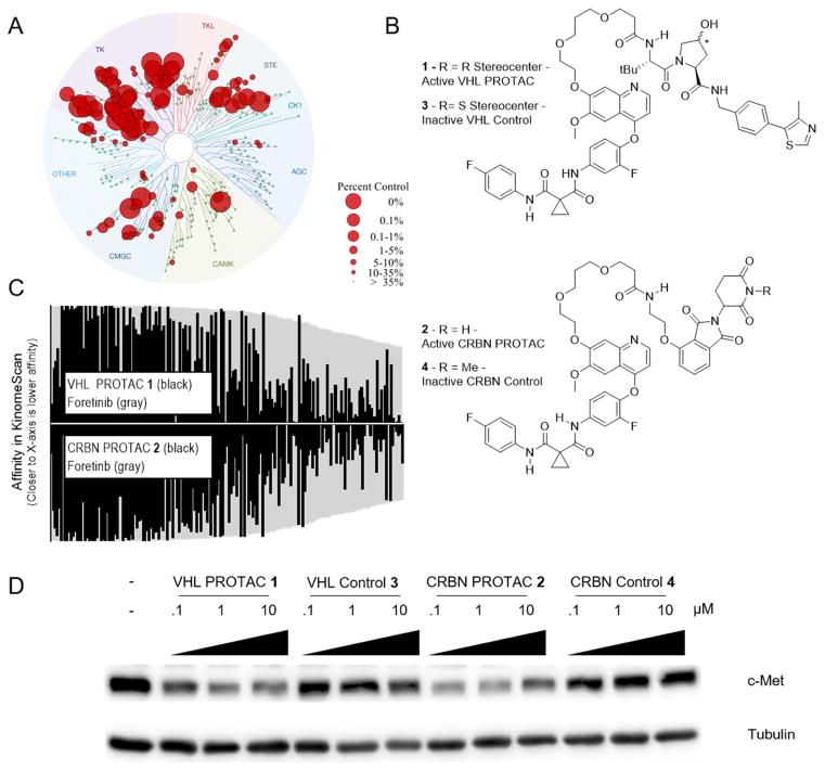
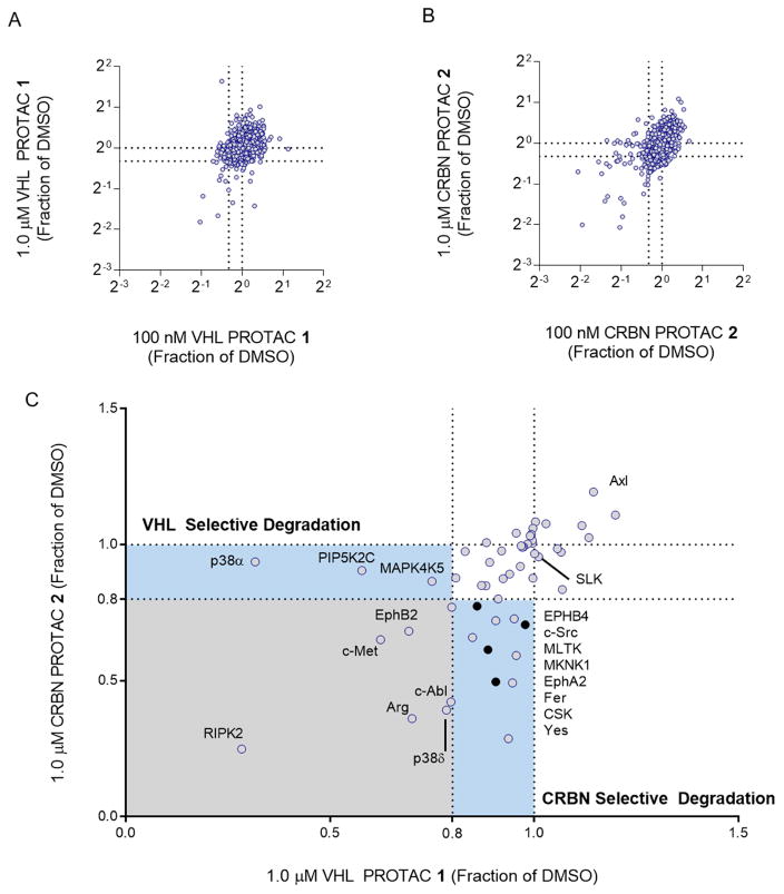
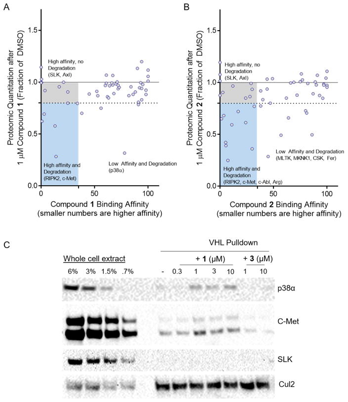
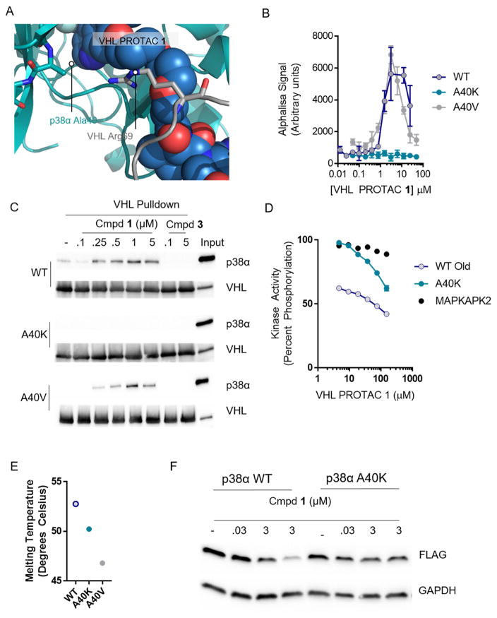
Inhibiting protein function selectively is a major goal of modern drug discovery. Here, we report a previously understudied benefit of small molecule proteolysis-targeting chimeras (PROTACs) that recruit E3 ubiquitin ligases to target proteins for their ubiquitination and subsequent proteasome-mediated degradation. Using promiscuous CRBN- and VHL-recruiting PROTACs that bind >50 kinases, we show that only a subset of bound targets is degraded. The basis of this selectivity relies on protein-protein interactions between the E3 ubiquitin ligase and the target protein, as illustrated by engaged proteins that are not degraded as a result of unstable ternary complexes with PROTAC-recruited E3 ligases. In contrast, weak PROTAC:target protein affinity can be stabilized by high-affinity target:PROTAC:ligase trimer interactions, leading to efficient degradation. This study highlights design guidelines for generating potent PROTACs as well as possibilities for degrading undruggable proteins immune to traditional small-molecule inhibitors.

Keywords: CRBN; MAPK/p38; PROTACs; VHL; chemical biology; protein degradation; protein-protein interactions; proteomics; selective degradation; ternary complex.


Figures

Figure 1
Figure 1. Foretinib PROTACs bind many kinases and potently degrade c-Met
(A) Foretinib binds to 133 kinases with a Percent of Control less than 35 as measured by a competitive binding assay. (B) Structures of the foretinib-based PROTACs used in this study. Top is the VHL-recruiting PROTAC compound 1. The stereocenter of the hydroxyproline VHL-binding element is R for the active PROTAC while it is S for the inactive control compound 3. Bottom, the Cereblon-recruiting PROTAC compound 2. The nitrogen atom of the glutarimide ring is non-methylated in the active PROTAC, while in the inactive control compound 4 it is methylated. (C) The affinity for most kinases changes upon addition of the linker and E3-ligase recruiting element. Along the X-axis are the 133 kinases that are considered hits for foretinib (i.e. having a percent of control value of 35 or less in KinomeScan data) sorted in order of decreasing affinity for foretinib. The values for foretinib are plotted in grey on both the positive and negative Y-axes. On the positive Y-axis, the values for compound 1 are plotted in black, while the negative X-axis has the values for compound 2. See Supplemental Table 1 and Figure S1 for full KinomeScan data sets. (D) MDA-MB-231 cells were treated with the indicated concentrations of compounds 1–4 for 24 hours, and c-Met protein levels were analyzed by immunoblot.
Figure 2
Figure 2. Foretinib-based PROTACs degrade only a subset of foretinib-binding kinases
(A) Multiplexed tandem mass spectrometry was used to assess global changes in the proteome of MDA-MB-231 cells after treatment with VHL PROTAC 1. After 24 hours with either 100 nM (plotted on the X-axis) or 1 μM (Y-axis) of 1, proteins were extracted from cells, labeled with tandem mass tags, and then 7,826 unique proteins were quantified according to the STAR methods section. The values plotted are fractions of the vehicle (DMSO) control. Dotted lines at 0.8 indicate the cut-off for a protein to be degraded. (B) Same as in (A), for CRBN PROTAC 2. (C) Comparison of proteomic changes after treatment with compounds 1 or 2. Fraction of DMSO after treatment with 1 μM VHL PROTAC 1 (X-Axis) or CRBN PROTAC 2 (Y-axis) are compared for each of the 54 foretinib-binding kinases. Dotted lines indicate the 80% cutoff for degradation, and areas of selectivity for 1 or 2 are highlighted. The light blue dots indicate proteins that are degraded in PROTAC treatment and in the control compounds, and so are not classified as bona fide PROTAC targets.
Figure 3
Figure 3. Affinity does not correlate with degradation efficiency, and stable ternary complexes more robustly predict degradation
(A) Comparison of VHL PROTAC 1’s degradation efficiency and binding affinity for 54 kinases. For each foretinib-binding kinase expressed in MDA-MB-231 cells, the fraction remaining by whole cell proteomics after treatment with 1 μM VHL PROTAC 1 is shown on the Y-axis as a fraction of the DMSO-treated samples. PROTAC’s affinity for that target is shown on the X-axis as a percent of control (KinomeScan). A target is considered degraded if it’s percent of DMSO is below 80%, and a target is considered to bind to the PROTAC if it’s percent of control is less than 35%. (B) Same as in (A), except for CRBN PROTAC 2. (C) Degraded kinases form stable ternary complexes with VHL. GST-tagged VHL/Elongin B/Elongin C were immobilized on glutathione beads and incubated with whole cell extract of MDA-MB-231 cells with the indicated concentrations of compounds 1 or 3. The beads were washed and bound proteins eluted with SDS buffer and analyzed by western blot. See Figure S2 for more robust characterization of degradation and affinity, and Figure S3C for further ternary complex blots of degraded targets.
Figure 4
Figure 4. Favorable protein-protein interactions stabilize the compound 1-induced complex between p38α and VHL
(A) VHL, PROTAC 1, and p38α were docked and a 120 ns molecular dynamics simulation relaxed the structure. Alanine 40 on p38α interacts with the kinked linker region of PROTAC 1. (B) Proximity-based AlphaLisa detects a ternary complex between GST-VHL and His-p38α. VHL PROTAC 1 and purified p38α and VHL were incubated in the presence of Glutathione Donor and Anti-his acceptor beads, and the extent of ternary complex formation was assessed by excitation at 680 nm and detection of emission at 615 nm. (C) Stable interaction between p38α and VHL are interrupted by the A40K mutation. Immobilized VHL/EloB/EloC was used as a bait to trap purified p38α in the presence of the indicated concentrations of compound 1 or 3. The far right lane represents a 1:25 dilution of the total protein used in each pulldown reaction. (D) Despite inhibition of ternary complex formation, the A40K mutation is inhibited by VHL PROTAC 1 to a similar extent as measured by a Z′Lyte cascade assay in which active p38α phosphorylates inactive MAPKAPK2 which phosphorylates a FRET-pair-containing peptide. Active MAPKAPK2 (without p38α protein) was also included as s control. (E) Thermal Stability of the WT, A40K, and A40 mutants are similar, as assessed by Sypro Orange fluorescence with increasing temperature. (F) Overexpressed wildtype p38α is degraded in cells, but not the A40K mutant. HeLa cells were transfected with a pcDNA5 vector containing FLAG-tagged p38α (wildtype or p38α), and then treated with VHL PROTAC 1 at the indicated concentrations for 24 hours.

Comment in

Similar articles

Cited by

References

    1. An J, Ponthier CM, Sack R, Seebacher J, Stadler MB, Donovan KA, Fischer ES. pSILAC mass spectrometry reveals ZFP91 as IMiD-dependent substrate of the CRL4CRBN ubiquitin ligase. Nat Commun. 2017;8:15398. - PMC - PubMed
    1. Bondeson DP, Mares A, Smith IED, Ko E, Campos S, Miah AH, Mulholland KE, Routly N, Buckley DL, Gustafson JL, et al. Catalytic in vivo protein knockdown by small-molecule PROTACs. Nat Chem Biol. 2015;11:611–617. - PMC - PubMed
    1. Buckley DL, Van Molle I, Gareiss PC, Tae HS, Michel J, Noblin DJ, Jorgensen WL, Ciulli A, Crews CM. Targeting the von Hippel–Lindau E3 Ubiquitin Ligase Using Small Molecules To Disrupt the VHL/HIF-1α Interaction. J Am Chem Soc. 2012a;134:4465–4468. - PMC - PubMed
    1. Buckley DL, Gustafson JL, Van Molle I, Roth AG, Tae HS, Gareiss PC, Jorgensen WL, Ciulli A, Crews CM. Small-molecule inhibitors of the interaction between the E3 ligase VHL and HIF1α. Angew Chem Int Ed Engl. 2012b;51:11463–11467. - PMC - PubMed
    1. Buckley DL, Raina K, Darricarrere N, Hines J, Gustafson JL, Smith IE, Miah AH, Harling JD, Crews CM. HaloPROTACS: Use of Small Molecule PROTACs to Induce Degradation of HaloTag Fusion Proteins. ACS Chem Biol. 2015;10:1831–1837. - PMC - PubMed

Publication types

MeSH terms

Substances