. 2017:2017:4064628.
doi: 10.1155/2017/4064628. Epub 2017 Oct 10.

Increasing the Fungicidal Action of Amphotericin B by Inhibiting the Nitric Oxide-Dependent Tolerance Pathway

Affiliations

Increasing the Fungicidal Action of Amphotericin B by Inhibiting the Nitric Oxide-Dependent Tolerance Pathway

Kim Vriens et al. Oxid Med Cell Longev. 2017.

Abstract

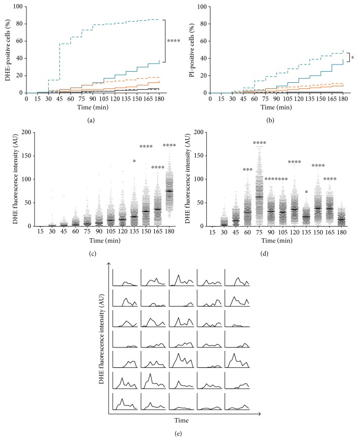
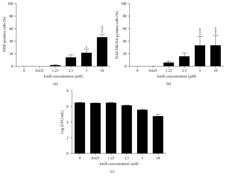
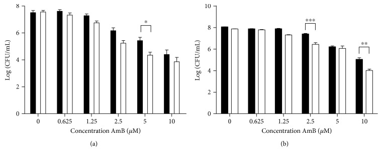
Amphotericin B (AmB) induces oxidative and nitrosative stresses, characterized by production of reactive oxygen and nitrogen species, in fungi. Yet, how these toxic species contribute to AmB-induced fungal cell death is unclear. We investigated the role of superoxide and nitric oxide radicals in AmB's fungicidal activity in Saccharomyces cerevisiae, using a digital microfluidic platform, which enabled monitoring individual cells at a spatiotemporal resolution, and plating assays. The nitric oxide synthase inhibitor L-NAME was used to interfere with nitric oxide radical production. L-NAME increased and accelerated AmB-induced accumulation of superoxide radicals, membrane permeabilization, and loss of proliferative capacity in S. cerevisiae. In contrast, the nitric oxide donor S-nitrosoglutathione inhibited AmB's action. Hence, superoxide radicals were important for AmB's fungicidal action, whereas nitric oxide radicals mediated tolerance towards AmB. Finally, also the human pathogens Candida albicans and Candida glabrata were more susceptible to AmB in the presence of L-NAME, pointing to the potential of AmB-L-NAME combination therapy to treat fungal infections.


Figures

Figure 1
Figure 1
AmB induced accumulation of superoxide and nitric oxide radicals and membrane permeabilization in S. cerevisiae. Yeast cultures were treated with different concentrations of AmB for 3 h and subjected to flow cytometry or plating assays. (a) Levels of superoxide radical detected by dihydroethidium (DHE) fluorescence and flow cytometry. (b) Levels of nitric oxide radical detected by 4-amino-5-methylamino-2′,7′-difluorofluorescein diacetate (DAF-FM DA) fluorescence and flow cytometry. (c) Membrane permeabilization events detected by propidium iodide (PI) fluorescence and flow cytometry. (d) Number of CFU/mL in Log-scale, assessed by plating assays and CFU counting. Means and standard errors of the means (SEM) of at least 3 independent biological experiments (n ≥ 3) are presented. Two-way ANOVA followed by Dunnett multiple comparison test was performed to analyse statistically significant differences in the number of PI-, DHE-, and DAF-FM DA-positive cells and cells able to proliferate between control treatment and treatment with different concentrations of AmB. ∗∗∗ and ∗∗∗∗ represent P < 0.001 and P < 0.0001, respectively.
Figure 2
Figure 2
AmB-induced superoxide radical accumulation, membrane permeabilization, and loss of proliferative capacity can be increased by blocking nitric oxide radical production using L-NAME. Exponential yeast cultures were treated with different concentrations of AmB in the presence or absence of 200 mM L-NAME for 3 h. (a) Levels of superoxide radical detected by dihydroethidium (DHE) fluorescence and flow cytometry. (b) Membrane permeabilization events detected by propidium iodide (PI) fluorescence and flow cytometry. (c) Number of CFU/mL in Log-scale, assessed by plating assays and CFU counting. Means and standard errors of the means (SEMs) of at least 3 independent biological experiments (n ≥ 3) are presented. Black bars represent treatment with AmB alone; white bars represent treatment with AmB supplemented with 200 mM L-NAME. Two-way ANOVA followed by Tukey multiple comparison test was performed to analyse significant differences between the two treatments. ∗ and ∗∗∗∗ represent P < 0.05 and P < 0.0001, respectively. Multiplicity-adjusted P values are presented in the text.
Figure 3
Figure 3
L-NAME increased and accelerated AmB-induced superoxide radical accumulation, membrane permeabilization, and intracellular superoxide radical levels. (a-b) Accumulation of superoxide radicals (a) and membrane permeabilization (b) in S. cerevisiae cells treated either with 0 μM (black), 5 μM (orange), or 10 μM (blue) AmB in the presence (dashed lines) or absence (solid lines) of 200 mM L-NAME during 3 h in 15 min intervals. Log-rank tests were performed to analyse significant differences between AmB treatment and treatment of AmB in combination with 200 mM L-NAME for each AmB dose. Data of at least 3 independent biological experiments is presented (n ≥ 3). ∗ and ∗∗∗∗ represent P < 0.05 and P < 0.0001, respectively. (c-d) Intracellular DHE fluorescence in S. cerevisiae cells treated with 10 μM AmB in the absence (c) or presence (d) of 200 mM L-NAME. Single cells were monitored for their DHE fluorescence during treatment for 3 h in 15 min intervals using fluorescence microscopy and the DMF platform. The fluorescence intensity of each cell is presented as arbitrary units (AU), and each dot represents a single cell. Means and standard errors of the means (SEMs) of at least 3 independent biological experiments (n ≥ 3), with at least 780 cells each, are presented. Two-way ANOVA followed by Tukey multiple comparison test was performed to analyse significant differences in DHE fluorescence intensity. ∗, ∗∗∗, and ∗∗∗∗ represent P < 0.05, P < 0.001, and P < 0.0001, respectively. (e) DHE fluorescence intensity of individual cells over time. A selection of 35 cells was randomly chosen and is representative for more than 3000 cells that were analysed in this study. Each plot represents the DHE fluorescence intensity, measured every 15 min, of one representative cell over the whole duration of the experiment, that is, 180 min.
Figure 4
Figure 4
L-NAME decreased the proliferative capacity of cells during AmB treatment, which seems independent of superoxide radical accumulation. Exponential yeast cells were treated with 10 μM AmB in the presence (white bars) or absence (black bars) of 200 mM L-NAME for 3 h. Cells were analysed for their DHE and PI fluorescence in the DMF setup (a and b) or subjected to bulk plating assays (c) every 15 min. Means and standard error of the means (SEMs) of at least 3 independent biological experiments (n ≥ 3) are presented. Two-way ANOVA followed by Tukey multiple comparison test was performed to analyse significant differences between the two treatments; Two-way ANOVA followed by Dunnett multiple comparison test was performed to analyse significant differences between the first data point (i.e., 0 min (in (c)) or 15 min (in (a) and (b))) and other data points within the same treatment (only the primary significant difference is presented to avoid overcrowding of the figure); ∗, ∗∗, and ∗∗∗∗ represent P < 0.05, P < 0.01, and P < 0.0001, respectively. A dotted line is shown at 15 min to point out the clear differences between the responses at this time point.
Figure 5
Figure 5
The nitric oxide donor, S-nitrosoglutathione, inhibited the killing activity of AmB. Yeast cells were treated with different concentrations of AmB, in the absence (black) or presence of 200 mM L-NAME (blue) or 2 mM S-nitrosoglutathione (orange). Means and standard errors of the means (SEMs) of at least two independent biological experiments (n ≥ 2) are presented. The number of CFU/mL for different treatments (insert) was assessed by plating assays and CFU counting and is shown relative to the number of CFU/mL at the start of the experiment. Two-way ANOVA followed by Tukey multiple comparison test was performed to analyse significant differences between the two treatments; ∗ and ∗∗∗∗ represent P < 0.05 and P < 0.0001, respectively.
Figure 6
Figure 6
Amphotericin B induced cell cycle arrest in the G2/M phase in yeast. Exponential yeast cultures were treated with either control (1% DMSO; 10% mQ), 200 mM L-NAME (dissolved in mQ), 10 μM AmB (dissolved in DMSO), or a combination of the above for 7.5 min (a) and 15 min (b). After treatment, cells were washed with PBS, fixed in 70% EtOH, stained with PI, and subjected to flow cytometry for cell cycle analysis. White bars represent cells in the G0/G1 phase, black bars represent cells in the S phase, and pixelated bars represent cells in the G2/M phase. Means and standard error of the means (SEMs) of 3 independent biological experiments (n = 3) are presented. Two-way ANOVA followed by Tukey multiple comparison test was performed to analyse differences between the cell cycle distributions of control treatment and AmB, L-NAME, or AmB + L-NAME treatment and between cell cycle distributions of AmB treatment and AmB + L-NAME treatment. ∗, ∗∗, ∗∗∗, and ∗∗∗∗ represent P < 0.05, P < 0.01, P < 0.001, and P < 0.0001, respectively. Multiplicity-adjusted P values are presented in the main text.
Figure 7
Figure 7
AmB induced accumulation of superoxide and nitric oxide radicals in C. albicans and decreased the number of cells that are able to proliferate. Exponential C. albicans cultures were treated with different concentrations of AmB for 3 h at room temperature and subjected to flow cytometry or plating assays. (a) Levels of superoxide radical detected by dihydroethidium (DHE) fluorescence and flow cytometry. (b) Levels of nitric oxide radical detected by 4-amino-5-methylamino-2′,7′-difluorofluorescein diacetate (DAF-FM DA) fluorescence and flow cytometry. (c) Number of CFU/mL in Log-scale, assessed by plating assays and CFU counting. Means and standard errors of the means (SEMs) of at least 3 independent biological experiments (n ≥ 3) are presented. Two-way ANOVA followed by Dunnett multiple comparison test was performed to analyse statistically significant differences in the number of DHE- and DAF-FM DA-positive cells and cells able to proliferate between control treatment and treatment with different concentrations of AmB. ∗ and ∗∗∗∗ represent P < 0.05 and P < 0.0001, respectively.
Figure 8
Figure 8
L-NAME significantly decreased the number of AmB-treated cells that are able to proliferate in C. albicans (a) and C. glabrata (b). Exponential C. albicans and C. glabrata cultures were treated with different dosages of AmB in the presence (white bars) or absence (black bars) of 200 mM L-NAME for 180 min at 37°C and subjected to plating assays. Means and standard errors of the means (SEMs) of 3 independent biological experiments (n = 3) are presented. Two-way ANOVA followed by Tukey multiple comparison test was performed to analyse significant differences between the two treatments; ∗, ∗∗, and ∗∗∗ represent P < 0.05, P < 0.01, and P < 0.001, respectively.
Figure 9
Figure 9
Schematic overview of the major findings on AmB mechanism of action in this study. Within 15 min, AmB caused cell cycle arrest in the G2/M phase and induced a yet to be elucidated event X, the latter leading to loss of proliferative capacity in yeast. These effects were independent of nitric oxide radicals, superoxide anion radicals, and membrane permeabilization. After 30 min, AmB induced the accumulation of superoxide radicals, which was associated with membrane permeabilization and loss of proliferative capacity in yeast, and was partially blocked by beneficial action of nitric oxide radicals. Interestingly, the combinatorial action of AmB and L-NAME induced a yet to be identified event Y within 15 min, which was independent of nitric oxide radicals, and enhanced the effect of event X, leading to enhanced loss of proliferative capacity in yeast.

Similar articles

Cited by

References

    1. Brown A. J., Haynes K., Quinn J. Nitrosative and oxidative stress responses in fungal pathogenicity. Current Opinion in Microbiology. 2009;12(4):384–391. doi: 10.1016/j.mib.2009.06.007. - DOI - PMC - PubMed
    1. Kaloriti D., Tillmann A., Cook E., et al. Combinatorial stresses kill pathogenic Candida species. Medical Mycology. 2012;50(7):699–709. doi: 10.3109/13693786.2012.672770. - DOI - PMC - PubMed
    1. Ferrari C. K., Souto P. C., Franca E. L., Honorio-Franca A. C. Oxidative and nitrosative stress on phagocytes’ function: from effective defense to immunity evasion mechanisms. Archivum Immunologiae et Therapiae Experimentalis. 2011;59(6):441–448. doi: 10.1007/s00005-011-0144-z. - DOI - PubMed
    1. Hromatka B. S., Noble S. M., Johnson A. D. Transcriptional response of Candida albicans to nitric oxide and the role of the YHB1 gene in nitrosative stress and virulence. Molecular Biology of the Cell. 2005;16(10):4814–4826. doi: 10.1091/mbc.E05-05-0435. - DOI - PMC - PubMed
    1. Moradas-Ferreira P., Costa V. Adaptive response of the yeast Saccharomyces cerevisiae to reactive oxygen species: defences, damage and death. Redox Report. 2000;5(5):277–285. doi: 10.1179/135100000101535816. - DOI - PubMed

MeSH terms

LinkOut - more resources