

Spontaneous Bilateral Sternoclavicular Joint Septic Arthritis and Lumbar Discitis: An Unusual Case in a Healthy Adult
- PMID: 29130010
- PMCID: PMC5654277
- DOI: 10.1155/2017/7101694
Spontaneous Bilateral Sternoclavicular Joint Septic Arthritis and Lumbar Discitis: An Unusual Case in a Healthy Adult
Abstract
Introduction: Septic arthritis of the sternoclavicular (SC) joint is a rare condition. Typically, it presents in patients with risk of infection and is usually unilateral. In this report, we describe a case of spontaneous bilateral sternoclavicular joint infection of an otherwise healthy adult.
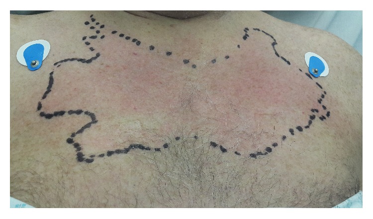
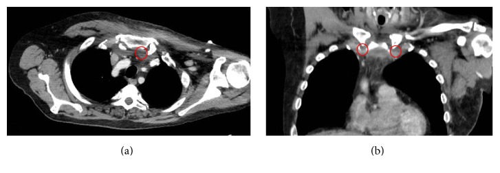
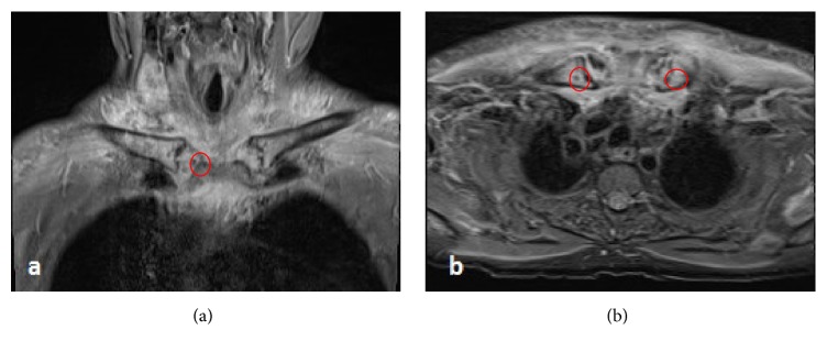
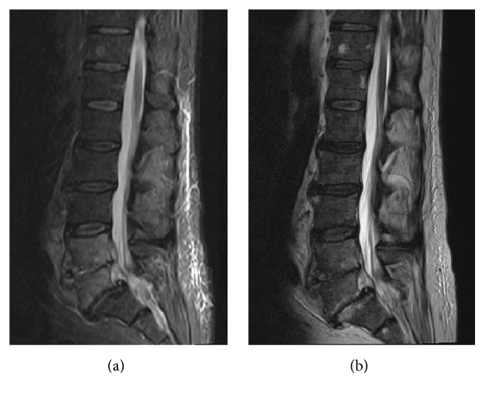
Case presentation: A 67-year-old man presented in our hospital complaining of 2-week history of neck and chest pain which was radiating to his shoulders bilaterally. Clinical examination revealed erythema and swelling of the sternoclavicular area. Inflammatory markers were raised. Image investigation with CT and MRI was undertaken and verified the presence of bilateral sternoclavicular joint infection. The patient received prolonged course of intravenous antibiotics since his admission. The patient was discharged in a good condition and followed up in clinic.
Conclusion: High index of clinical suspicion of SC joint infection is important for early diagnosis to avoid further complications.
Figures
Similar articles
-
Arthritis of the sternoclavicular joint masquerading as rupture of the cervical oesophagus: a case report.J Med Case Rep. 2009 Jan 29;3:40. doi: 10.1186/1752-1947-3-40. J Med Case Rep. 2009. PMID: 19178739 Free PMC article.
-
Unilateral sternoclavicular arthritis: inflammatory arthritis or septic arthritis, that is the question - a case report.J Int Med Res. 2022 Apr;50(4):3000605221089786. doi: 10.1177/03000605221089786. J Int Med Res. 2022. PMID: 35387512 Free PMC article.
-
Sternoclavicular Septic Arthritis: A Case Report.Cureus. 2023 Apr 25;15(4):e38130. doi: 10.7759/cureus.38130. eCollection 2023 Apr. Cureus. 2023. PMID: 37252575 Free PMC article.
-
Septic arthritis of the lumbar facet joint presenting as spontaneous bacterial peritonitis: a rare case requiring surgical intervention.Eur J Orthop Surg Traumatol. 2020 Jan;30(1):175-178. doi: 10.1007/s00590-019-02527-y. Epub 2019 Aug 29. Eur J Orthop Surg Traumatol. 2020. PMID: 31463672 Review.
-
Septic arthritis of a lumbar facet joint and a sternoclavicular joint.Spine (Phila Pa 1976). 1995 Jun 1;20(11):1304-6. doi: 10.1097/00007632-199506000-00021. Spine (Phila Pa 1976). 1995. PMID: 7660242 Review.
Cited by
-
Salmonella Sternoclavicular Septic Arthritis in a Non-sickle Cell Disease Patient.Cureus. 2023 Jan 23;15(1):e34094. doi: 10.7759/cureus.34094. eCollection 2023 Jan. Cureus. 2023. PMID: 36843725 Free PMC article.
-
Diagnosis and management of sternoclavicular joint infections: a literature review.J Thorac Dis. 2020 Aug;12(8):4418-4426. doi: 10.21037/jtd-20-761. J Thorac Dis. 2020. PMID: 32944355 Free PMC article. Review.
-
Acute Sternoclavicular Joint Sepsis With Medial Clavicle Osteomyelitis (Staphylococcus aureus) and Cervical-Thoracic Epidural Phlegmon in an Adult Female With No Apparent Risk Factors.Cureus. 2023 Mar 7;15(3):e35870. doi: 10.7759/cureus.35870. eCollection 2023 Mar. Cureus. 2023. PMID: 37033534 Free PMC article.
References
-
- Blankstein A., Nerubay J., Lin E., Keren G., Friedman B., Horoszowski H. Septic arthritis of the sternoclavicular joint. Orthopaedic Review. 1986;15(7):440–442. - PubMed
Publication types
LinkOut - more resources
Full Text Sources
Other Literature Sources
