. 2017:2017:7868645.
doi: 10.1155/2017/7868645. Epub 2017 Oct 10.

The PDZ-Binding Motif of HPV16-E6 Oncoprotein Modulates the Keratinization and Stemness Transcriptional Profile In Vivo

Affiliations

The PDZ-Binding Motif of HPV16-E6 Oncoprotein Modulates the Keratinization and Stemness Transcriptional Profile In Vivo

Enoc Mariano Cortés-Malagón et al. Biomed Res Int. 2017.

Abstract

Objective: The aim of this work was to compare the early gene expression profiles in the skin of HPV16-E6 transgenic mice regulated by the E6 PDZ-binding motif.

Materials and methods: The global transcriptional profiles in dorsal skin biopsies from K14E6 and K14E6Δ146-151 transgenic mice were compared using microarrays. Relevant genes obtained from the most differentially expressed processes were further examined by RT-qPCR, in situ RT-PCR, Western blot, or immunofluorescence.

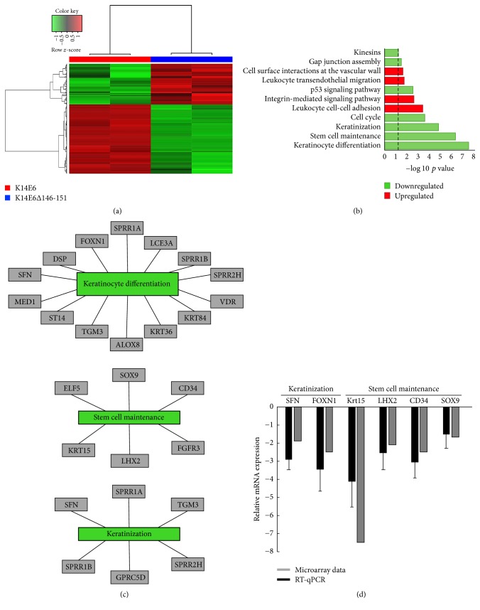
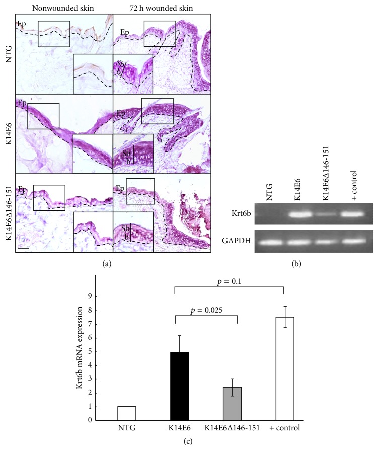
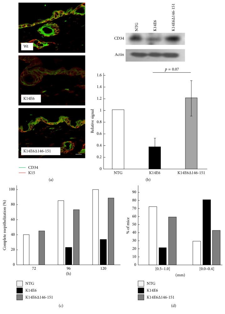
Results: The transcriptomic landscape of K14E6 versus K14E6Δ146-151 shows that the most affected expression profiles were those related to keratinocyte differentiation, stem cell maintenance, and keratinization. Additionally, downregulation of epidermal stemness markers such as K15 and CD34, as well as the upregulation of cytokeratin 6b, appeared to be dependent on the E6 PDZ-binding motif. Finally, wound healing, a physiological process linked to stemness, is impaired in the K14E6 mice compared to K14E6Δ146-151.

Conclusion: The E6 PDZ-binding motif appears to affect stemness and keratinization during early stages of skin carcinogenesis. As E6 plays a significant role in HPV-induced skin carcinogenesis, the K14E6 versus K14E6Δ146-151 transcriptional profile provides a source of valuable data to uncover novel E6 functions in the skin.


Figures

Figure 1
Figure 1
Global transcriptional analysis of K14E6 versus K14E6Δ146-151 mice. (a) Hierarchical clustering of genes differentially expressed in an RNA-microarray analysis performed on K14E6 versus K14E6Δ146-151 comparison. A z-score adjusts heatmap. (b) Pathway enrichment plot of differentially expressed genes. The plot shows the significantly (−log⁡10  p value) upregulated (red bars) and downregulated (green bars) pathways of differentially expressed genes. (c) Networks of three of the most negatively enriched cellular processes (keratinocyte differentiation, stem cell maintenance, and keratinization) based on statistical significance (−log⁡10  p value). (d) Microarray validation by RT-qPCR. Grey bars represent microarray expression value versus black bars which represent RT-qPCR validation; each RT-qPCR validation was performed as n = 3 biological replicates.
Figure 2
Figure 2
Expression and cellular localisation of keratin 6b mRNA. (a) In situ RT-PCR showing the cellular localisation of mRNA in nonwounded, and 72 h wounded dorsal skin evaluated in 1.5-month-old NTG, K14E6, and K14E6Δ146-151 transgenic mice. Magnification: 20x. Dashed line: basal membrane. Ep: epidermis, Sb: suprabasal stratum; and b: basal stratum. (b) End-point RT-PCR showing the amplification products of K6b and GAPDH genes. (c) The RT-qPCR assay is showing the relative expression of Krt6 using the amount of target = 2−ΔΔCt equation; Krt6 RT-qPCR was performed as n = 3 biological replicates. Positive control for (b) and (c) refers to 72 h wounded skin samples. Magnification: 20x.
Figure 3
Figure 3
E-Cadherin and β-catenin cellular localisation in the skin. Immunofluorescence assay of nonwounded skin showing the expression and cellular localisation of E-cadherin (FITC-signal) or β-catenin (TRITC-signal) proteins in 1.5-month-old NTG, K14E6, and K14E6Δ146-151 transgenic mice. Insets highlight the nuclear signal indicated by arrows. Magnification: 40x.
Figure 4
Figure 4
CD34/Krt15 epidermal stemness markers and reepithelization/wound closure assays. (a) Immunofluorescence assay of nonwounded skin showing the expression and colocalization of CD34 (FITC-signal) and K5 (TRITC-signal) stemness markers. (b) Western blot of CD34 evaluated in total protein extracts of nonwounded skin samples. The graph shows the result of n = 4 independent biological replicates. (c) Epithelization assay performed in 72, 96, and 120 h wounded dorsal skin indicating the percentage of complete epithelization for each mice strain. (d) Wound closure measure 28 days after wounding evaluated on ears. The hole diameter was measured with a micro ruler using a stereoscopic microscope. Magnification of (a): 40x.

Similar articles

Cited by

References

    1. zur Hausen H. Papillomaviruses and cancer: from basic studies to clinical application. Nature Reviews Cancer. 2002;2(5):342–350. doi: 10.1038/nrc798. - DOI - PubMed
    1. Walboomers J. M. M., Jacobs M. V., Manos M. M., et al. Human papillomavirus is a necessary cause of invasive cervical cancer worldwide. The Journal of Pathology. 1999;189(1):12–19. doi: 10.1002/(sici)1096-9896(199909)189:1lt;12::aid-path431x0003e;3.0.co;2-f. - DOI - PubMed
    1. Nguyen H. P., Ramírez-Fort M. K., Rady P. L. The biology of human papillomaviruses. Current Problems in Dermatology (Switzerland) 2014;45:19–32. doi: 10.1159/000355959. - DOI - PubMed
    1. Pfister H. Chapter 8: Human papillomavirus and skin cancer. Journal of the National Cancer Institute. Monographs. 2003;(31):52–56. - PubMed
    1. Andersson K., Luostarinen T., Strand A. S., et al. Prospective study of genital human papillomaviruses and nonmelanoma skin cancer. International Journal of Cancer. 2013;133(8):1840–1845. doi: 10.1002/ijc.28188. - DOI - PubMed

MeSH terms