. 2017 Dec;38(6):3355-3366.
doi: 10.3892/or.2017.6052. Epub 2017 Oct 23.

c-Fos mediates α1, 2-fucosyltransferase 1 and Lewis y expression in response to TGF-β1 in ovarian cancer

Affiliations

c-Fos mediates α1, 2-fucosyltransferase 1 and Lewis y expression in response to TGF-β1 in ovarian cancer

Yingying Hao et al. Oncol Rep. 2017 Dec.

Abstract

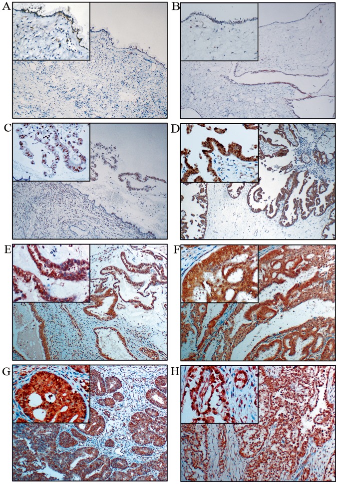
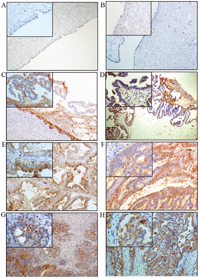
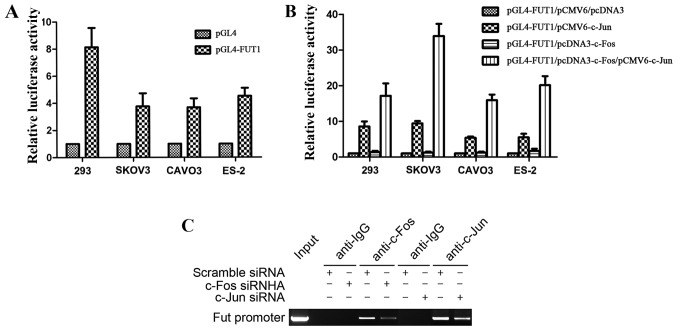
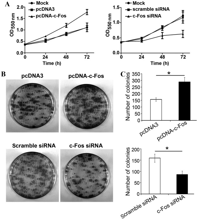
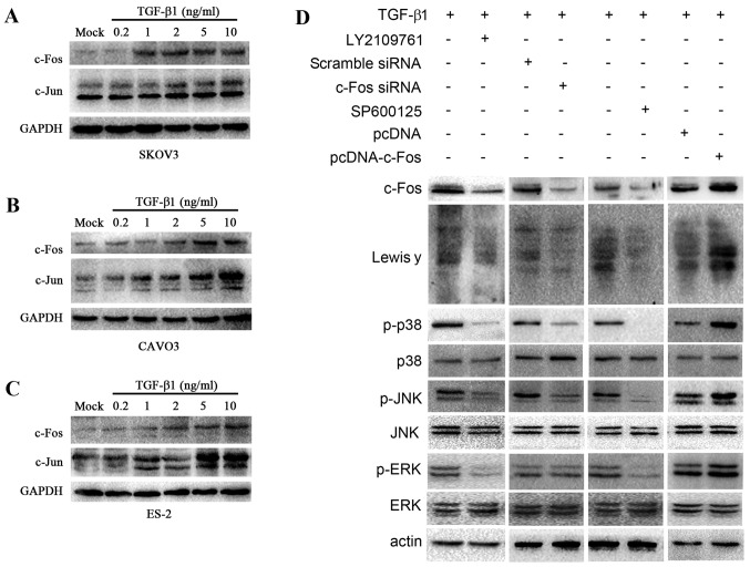
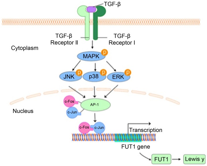
FUT1 is a key rate-limiting enzyme in the synthesis of Lewis y, a membrane-associated carbohydrate antigen. The aberrant upregulation of FUT1 and Lewis y antigen is related to proliferation, invasion and prognosis in malignant epithelial tumors. A c-Fos/activator protein-1 (AP-1) binding site was found in the FUT1 promoter. However, the mechanisms of transcriptional regulation of FUT1 remain poorly understood. TGF-β1 is positively correlated to Lewis y. In the present study, we investigated the molecular mechanism of FUT1 gene expression in response to TGF-β1. We demonstrated that c-Fos was highly expressed in 77.50% of ovarian epithelial carcinoma cases and was significantly correlated with Lewis y. Using luciferase activity and chromatin immunoprecipitation (ChIP) assay, we further revealed that c-Fos interacted with the FUT1 promoter in ovarian cancer cells and transcriptional capacity of the heterodimer formed by c-Fos and c-Jun was stronger than that of the c-Fos or c-Jun homodimers. Then, we demonstrated that TGF-β1 induced dose-dependent c-Fos expression, which was involved in TGF-β1-induced ovarian cancer cell proliferation. In addition, inhibition of MAPK activation or TGF-β1 receptor by pharmacological agents prevented TGF-β1-induced c-Fos and Lewis y expression. Silencing of c-Fos prevented TGF-β1-induced Lewis y expression. Collectively, the results of these studies demonstrated that TGF-β1 regulated FUT1 and Lewis y expression by activating the MAPK/c-Fos pathway.


Figures

Figure 1.
Figure 1.
Immunohistochemical staining of c-Fos in ovarian tumors. (A) Mucinous, (B) serous, (C) borderline mucinous and (D) borderline serous cystadenoma. (E) Mucinous cystadenocarcinoma and (F) serous cystadenocarcinoma. (G) Endometrioid and (H) clear cell carcinoma. (Original magnification ×100 and ×400 for the small box at the top-left corner).
Figure 2.
Figure 2.
Immunohistochemical staining of Lewis y in ovarian tumors. (A) Mucinous, (B) serous, (C) borderline mucinous and (D) borderline serous cystadenoma. (E) Mucinous cystadenocarcinoma and (F) serous cystadenocarcinoma. (G) Endometrioid carcinoma and (H) clear cell carcinoma (original magnification ×100 and ×400 for the small box at the top-left corner).
Figure 3.
Figure 3.
Role of c-Fos in the regulation of FUT1. (A) The activity of human FUT1 promoter construct in 293 cells and ovarian cancer cells lines was detected. (B) The luciferase reporter assay of FUT1 promoter constructs in 293 cells and ovarian cancer cells. Cell lines 293, SKOV3, CAVO3 and ES-2 were co-transfected with pGL4-FUT1, with empty vectors, or pCMV6-c-Jun or pcDNA3-c-Fos or both. (C) ChIP assay detected c-Fos and c-Jun interacted with FUT1 promoter in CAVO3 cells. Data represent the mean ± SD of 3 independent experiments performed in triplicate; *P<0.05.
Figure 4.
Figure 4.
TGF-β1 promotes cell proliferation via c-Fos in ovarian cancer cells. (A) Cell viability was detected using MTT cell proliferation assay in CAVO3 cells. CAVO3 cells were transfected with pcDNA3-c-Fos, pcDNA3 or c-Fos siRNA for 24 h, followed by pretreatment with TGF-β1. (B and C) Colony formation assessed cell proliferation ability. The cells and processing were the same as shown in a. The data are expressed as the mean ± SD of 3 independent experiments; *P<0.05, compared with the control.
Figure 5.
Figure 5.
TGF-β1 activates c-Fos through the MAPK/AP-1 pathway. Upon TGF-β1 treatment, western blotting was employed to determine c-Jun and c-Fos expression in 3 ovarian cells. (A) SKOV3, (B) CAVO3, (C) ES-2. c-Fos and c-Jun expression is related to TGF-β1 in a concentration manner in ovarian cells pretreated with various concentrations of TGF-β1 (0.2, 1, 2, 5 and 10 ng/ml) for 5 min. (D) Western blot analysis was carried out to determine the expression of c-Fos, Lewis y and the downstream signal elements of the MAPK signaling pathway, JNK, p38 and ERK, and the change in the phosphorylation level of JNK, p38 and ERK. CAVO3 cells were treated with pcDNA-c-Fos, c-Fos siRNA, inhibitor of TGF-β1 receptor (LY2109761) and JNK (SPF600125) upon TGF-β1 stimulation.
Figure 6.
Figure 6.
Pathway of c-Fos-mediated Lewis y in response to TGF-β1. TGF-β1 activates c-Fos/AP-1 through the MAPK pathway. As a transcriptional factor, c-Fos together with c-Jun upregulated FUT1 expression by binding to its promoter, consequently leading to the enhanced synthesis of Lewis y.

References

    1. Siegel R, Naishadham D, Jemal A. Cancer statistics, 2013. CA Cancer J Clin. 2013;63:11–30. doi: 10.3322/caac.21166. - DOI - PubMed
    1. Salani R, Bristow RE. Surgical management of epithelial ovarian cancer. Clin Obstet Gynecol. 2012;55:75–95. doi: 10.1097/GRF.0b013e31824b4629. - DOI - PubMed
    1. Coleman RL, Monk BJ, Sood AK, Herzog TJ. Latest research and treatment of advanced-stage epithelial ovarian cancer. Nat Rev Clin Oncol. 2013;10:211–224. doi: 10.1038/nrclinonc.2013.5. - DOI - PMC - PubMed
    1. Kaczmarek R. Alterations of Lewis histo-blood group antigen expression in cancer cells. Postepy Hig Med Dosw (Online) 2010;64:87–99. (In Polish) - PubMed
    1. Iwamori M, Iwamori Y, Kubushiro K, Ishiwata I, Kiguchi K. Characteristic expression of Lewis-antigenic glycolipids in human ovarian carcinoma-derived cells with anticancer drug-resistance. J Biochem. 2007;141:309–317. doi: 10.1093/jb/mvm031. - DOI - PubMed

MeSH terms