Review
. 2019 Apr 1;30(10):1331-1351.
doi: 10.1089/ars.2017.7403. Epub 2018 Jan 10.

Protein S-Nitrosylation: Determinants of Specificity and Enzymatic Regulation of S-Nitrosothiol-Based Signaling

Affiliations
Review

Protein S-Nitrosylation: Determinants of Specificity and Enzymatic Regulation of S-Nitrosothiol-Based Signaling

Colin T Stomberski et al. Antioxid Redox Signal. .

Abstract

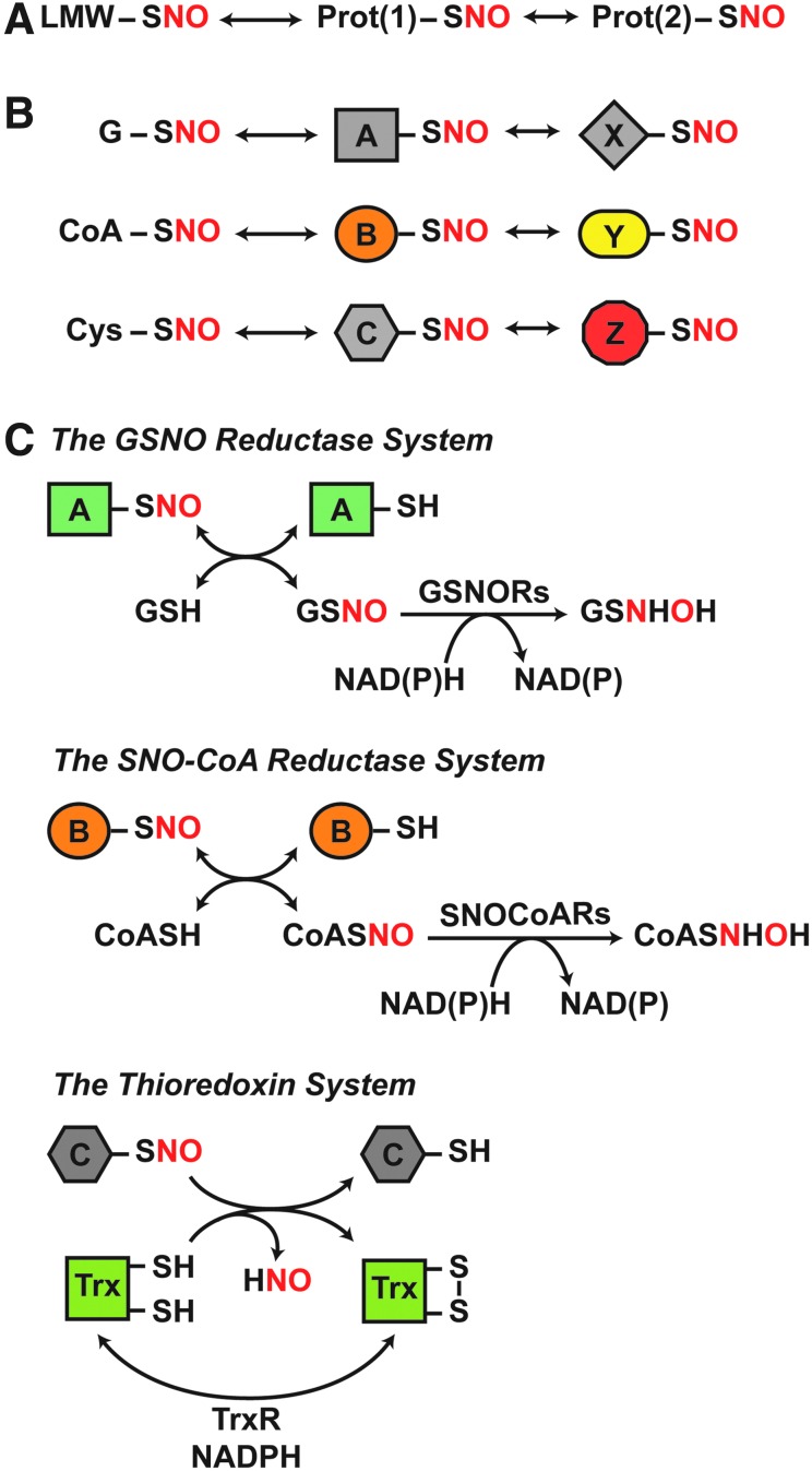
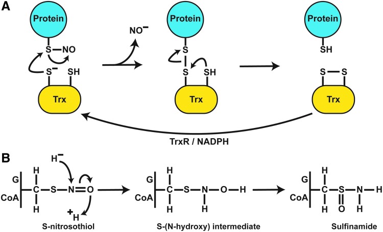
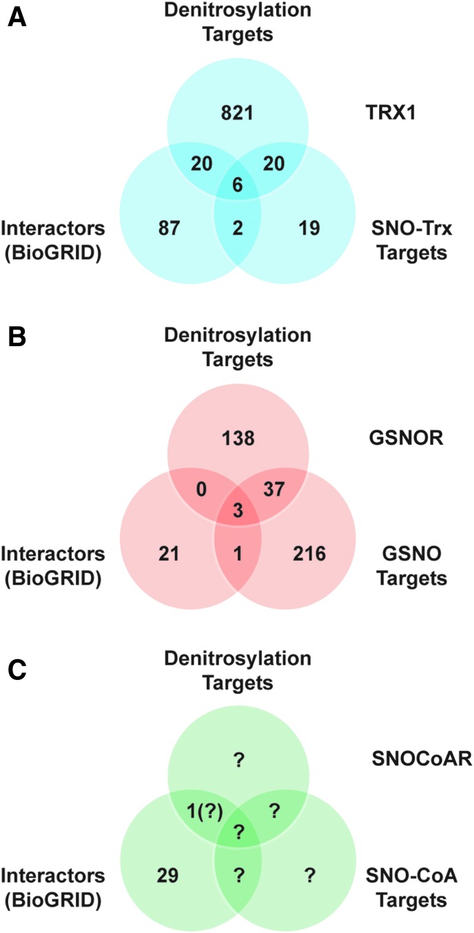
Significance: Protein S-nitrosylation, the oxidative modification of cysteine by nitric oxide (NO) to form protein S-nitrosothiols (SNOs), mediates redox-based signaling that conveys, in large part, the ubiquitous influence of NO on cellular function. S-nitrosylation regulates protein activity, stability, localization, and protein-protein interactions across myriad physiological processes, and aberrant S-nitrosylation is associated with diverse pathophysiologies. Recent Advances: It is recently recognized that S-nitrosylation endows S-nitroso-protein (SNO-proteins) with S-nitrosylase activity, that is, the potential to trans-S-nitrosylate additional proteins, thereby propagating SNO-based signals, analogous to kinase-mediated signaling cascades. In addition, it is increasingly appreciated that cellular S-nitrosylation is governed by dynamically coupled equilibria between SNO-proteins and low-molecular-weight SNOs, which are controlled by a growing set of enzymatic denitrosylases comprising two main classes (high and low molecular weight). S-nitrosylases and denitrosylases, which together control steady-state SNO levels, may be identified with distinct physiology and pathophysiology ranging from cardiovascular and respiratory disorders to neurodegeneration and cancer.

Critical issues: The target specificity of protein S-nitrosylation and the stability and reactivity of protein SNOs are determined substantially by enzymatic machinery comprising highly conserved transnitrosylases and denitrosylases. Understanding the differential functionality of SNO-regulatory enzymes is essential, and is amenable to genetic and pharmacological analyses, read out as perturbation of specific equilibria within the SNO circuitry.

Future directions: The emerging picture of NO biology entails equilibria among potentially thousands of different SNOs, governed by denitrosylases and nitrosylases. Thus, to elucidate the operation and consequences of S-nitrosylation in cellular contexts, studies should consider the roles of SNO-proteins as both targets and transducers of S-nitrosylation, functioning according to enzymatically governed equilibria.

Keywords: S-nitrosylase; S-nitrosylation; denitrosylation; nitric oxide; redox signaling.


Figures

<b>FIG. 1.</b>
FIG. 1.
Coupled, dynamic equilibria that govern protein S-nitrosylation are regulated by enzymatic denitrosylases. (A) SNO-proteins are in equilibrium with LMW-SNOs and can further participate in protein-to-protein transfer of the NO group (trans-S-nitrosylation) to subserve NO-based signaling. (B) Transnitrosylation by both identified LMW-SNOs (G, glutathione; CoA, coenzyme A; Cys, cysteine) and SNO-proteins will result in distinct sets of SNO-proteins that mediate specific SNO signaling cascades. (C) Distinct enzymatic denitrosylases regulate the coupled equilibria that confer specificity to SNO-based signaling. These include GSNORs and SNO-CoA reductases, which regulate protein S-nitrosylation by GSNO and SNO-CoA, respectively. These LMW-SNOs are in equilibrium with cognate SNO-proteins. In contrast, Trxs directly denitrosylate SNO-proteins. The reaction schemes illustrated are detailed in the Enzymatic Denitrosylation section. GSNO, S-nitrosoglutathione; GSNORs, GSNO reductases; LMW-SNOs, low-molecular-weight S-nitrosothiol; NO, nitric oxide; SNO, S-nitrosothiol; SNO-CoA, S-nitroso-coenzyme A; SNO-protein, S-nitroso-protein; Trx, thioredoxin.
<b>FIG. 2.</b>
FIG. 2.
Steady-state protein S-nitrosylation reflects denitrosylase activity. (A) In cultured human embryonic kidney cells, suppression of Trx-mediated denitrosylation with the TrxR inhibitor auranofin results in greatly enhanced steady-state levels of SNO-proteins (as detected by the SNO-RAC method) (57). (B) After induction of iNOS by systemic administration of bacterial lipopolysaccharide (LPS), steady-state levels of hepatic SNO-protein are greatly increased in the genetic absence of GSNOR [as assessed by photolysis-chemiluminescence; modified from Liu et al. (106)]. iNOS, inducible nitric oxide synthase; TrxR, thioredoxin reductase.
<b>FIG. 3.</b>
FIG. 3.
Differential RSNO reactivity. By default (middle scheme), a partial negative charge exists on the S atom of the SNO bond; a partial positive charge exists on the N atom. This intrinsically favors transnitrosylative reactions. Positive charge coordination with the S atom or negative charge coordination with the N or O atom would further increase electrophilicity of the N atom, enhancing the propensity for transnitrosylation (top). In contrast, negative charge coordination with the S atom or positive charge coordination with the N or O atom would increase electrophilicity of the S atom, preferencing S-thiolation reactions (bottom).
<b>FIG. 4.</b>
FIG. 4.
Enzymatic mechanisms of protein denitrosylation. (A) Denitrosylation by Trx requires a direct interaction with target SNO-proteins. Nucleophilic attack by an active Cys leads to mixed disulfide formation between Trx and target proteins with liberation of NO as a nitroxyl anion (likely as HNO). The mixed disulfide is resolved by the second active site Cys of Trx, generating oxidized Trx and reduced target protein thiol. Trx is subsequently reduced by TrxR and NADPH to regenerate denitrosylating activity. (B) A common reaction scheme exists for the reduction of GSNO and SNO-CoA by their cognate denitrosylases (GSNOR and SNO-CoA reductase). Hydride transfer from NAD(P)H to the N atom and protonation of the O atom lead to an S-(N-hydroxy) intermediate that rearranges to sulfinamide. HNO, nitroxyl.
<b>FIG. 5.</b>
FIG. 5.
Stimulus-coupled S-nitrosylation and denitrosylation: cardiomyocytes as an exemplary case. (A) Ligand-induced activation of the β2-AR in cardiomyocytes stimulates eNOS activity. eNOS-dependent S-nitrosylation of GRK2 suppresses GRK2 activity, whereas S-nitrosylation of β-arr2 facilitates β-arr2 activity, thereby regulating receptor desensitization and internalization. GSNOR negatively regulates the S-nitrosylation status of both GRK2 and β-arr2. (B) nNOS and GSNOR interact directly at the SR in cardiomyocytes. β2-AR activation stimulates nNOS-dependent S-nitrosylation of components of calcium handling (PLN, SERCA2a, RyR2) and of cardiac myofilaments, which is regulated by GSNOR-dependent denitrosylation. (A, B) SNO depicted in red indicates inhibition of normal function by S-nitrosylation; SNO depicted in green indicates activation of normal function by S-nitrosylation. Additional details are provided in the subsection “GSNOR in physiology and pathophysiology.” β-arr2, β-arrestin 2; β2-AR, beta-2 adrenergic receptor; eNOS, endothelial nitric oxide synthase; GRK2, G-protein-coupled receptor kinase 2; nNOS, neuronal nitric oxide synthase; PLN, phospholamban; RyR2, ryanodine receptor 2; SERCA2a, sarcoplasmic/endoplasmic reticulum calcium ATPase 2; SR, sarcoplasmic reticulum.
<b>FIG. 6.</b>
FIG. 6.
Proteinprotein interactions mediate enzymatic S-nitrosylation/denitrosylation. (A) For the Trx1 system, a Venn diagram shows overlap between the sets of interacting proteins, targets of denitrosylation, and targets of trans-S-nitrosylation. Interacting proteins were retrieved from BioGRID. Targets of Trx1 denitrosylation were obtained from Ben-Lulu et al. (17, 18). Targets of transnitrosylation by SNO-Trx1 are from Wu et al. (201). (B) A similar analysis for GSNOR also reveals overlap between interactors and targets of GSNOR-regulated denitrosylation, although the data sets are substantially smaller than in the case of Trx1. Targets of GSNOR-regulated denitrosylation are unique proteins from references in Table 2. Targets of GSNO-mediated S-nitrosylation are from Murray et al. (127) and Paige et al. (138). (C) Relatively few mammalian substrates of S-nitrosylation by SNO-CoA or of denitrosylation by SNO-CoAR have been identified to date, as indicated by question marks. SNO-CoAR, S-nitroso-coenzyme A reductase.

References

    1. Abunimer A, Smith K, Wu TJ, Lam P, Simonyan V, and Mazumder R. Single-nucleotide variations in cardiac arrhythmias: prospects for genomics and proteomics based biomarker discovery and diagnostics. Genes (Basel) 5: 254–269, 2014 - PMC - PubMed
    1. Anand P, Hausladen A, Wang Y, Zhang G, Stomberski C, Brunengraber H, Hess DT, and Stamler JS. Identification of S-nitroso-CoA reductases that regulate protein S-nitrosylation. Proc Natl Acad Sci U S A 111: 18572–18577, 2014 - PMC - PubMed
    1. Anand P. and Stamler JS. Enzymatic mechanisms regulating protein S-nitrosylation: implications in health and disease. J Mol Med 90: 233–244, 2012 - PMC - PubMed
    1. Andre FR, dos Santos PF, and Rando DG. Theoretical studies of the role of C-terminal cysteines in the process of S-nitrosylation of human Src kinases. J Mol Model 22: 23, 2016 - PubMed
    1. Angelo M, Singel DJ, and Stamler JS. An S-nitrosothiol (SNO) synthase function of hemoglobin that utilizes nitrite as a substrate. Proc Natl Acad Sci U S A 103: 8366–8371, 2006 - PMC - PubMed

LinkOut - more resources