

Evaluation of renal metabolic response to partial ureteral obstruction with hyperpolarized 13 C MRI
- PMID: 29130537
- PMCID: PMC5736002
- DOI: 10.1002/nbm.3846
Evaluation of renal metabolic response to partial ureteral obstruction with hyperpolarized 13 C MRI
Abstract
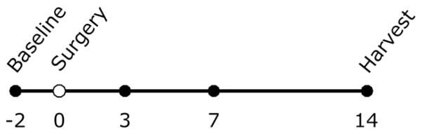
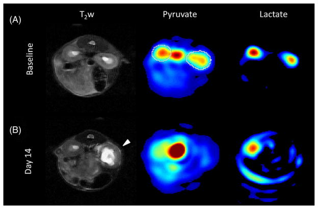
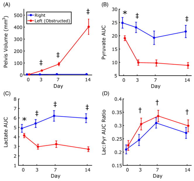
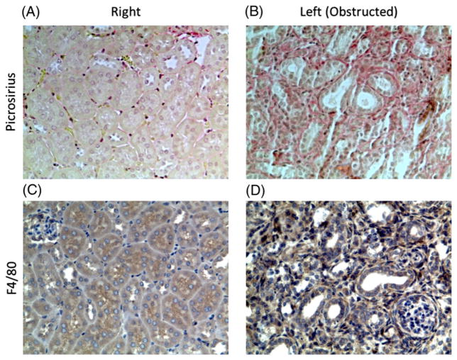
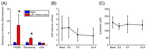
Hyperpolarized 13 C magnetic resonance imaging (MRI) may be used to non-invasively image the transport and chemical conversion of 13 C-labeled compounds in vivo. In this study, we utilize hyperpolarized 13 C MRI to evaluate metabolic markers in the kidneys longitudinally in a mouse model of partial unilateral ureteral obstruction (pUUO). Partial obstruction was surgically induced in the left ureter of nine adult mice, leaving the right ureter as a control. 1 H and hyperpolarized [1-13 C]pyruvate MRI of the kidneys was performed 2 days prior to surgery (baseline) and at 3, 7 and 14 days post-surgery. Images were evaluated for changes in renal pelvis volume, pyruvate, lactate and the lactate to pyruvate ratio. After 14 days, mice were sacrificed and immunohistological staining of both kidneys for collagen fibrosis (picrosirius red) and macrophage infiltration (F4/80) was performed. Statistical analysis was performed using a linear mixed effects model. Significant kidney × time interaction effects were observed for both lactate and pyruvate, indicating that these markers changed differently between time points for the obstructed and unobstructed kidneys. Both kidneys showed an increase in the lactate to pyruvate ratio after obstruction, suggesting a shift towards glycolytic metabolism. These changes were accompanied by marked hydronephrosis, fibrosis and macrophage infiltration in the obstructed kidney, but not in the unobstructed kidney. Our results show that pUUO is associated with increased pyruvate to lactate metabolism in both kidneys, with injury and inflammation specific to the obstructed kidney. The work also demonstrates the feasibility of the use of hyperpolarized 13 C MRI to study metabolism in renal disease.
Keywords: hyperpolarized 13C; metabolism; renal; ureteral obstruction.
Copyright © 2017 John Wiley & Sons, Ltd.
Figures
References
-
- [Accessed October 12, 2016];North American pediatric renal trials and collaborative studies annual report. 2014 https://web.emmes.com/study/ped/index.htm.
-
- Chevalier RL, Forbes MS, Thornhill BA. Ureteral obstruction as a model of renal interstitial fibrosis and obstructive nephropathy. Kidney Int. 2009;75(11):1145–1152. - PubMed
-
- Yoshioka Y, Tsutsumi T, Adachi M, Tokumura A. Altered phospholipid profile in urine of rats with unilateral ureteral obstruction. Metabolomics. 2009;5(4):429–433.
-
- MacLellan DL, Mataija D, Doucette A, et al. Alterations in urinary metabolites due to unilateral ureteral obstruction in a rodent model. Mol Biosyst. 2011;7(7):2181–2188. - PubMed
-
- Zhang H, Jia J, Cheng J, Ye F, Li X, Gao H. 1H NMR-based metabonomics study on serum of renal interstitial fibrosis rats induced by unilateral ureteral obstruction. Mol Biosyst. 2012;8(2):595–601. - PubMed
MeSH terms
Substances
Grants and funding
LinkOut - more resources
Full Text Sources
Other Literature Sources
Medical
