. 2017 Dec 1;127(12):4516-4526.
doi: 10.1172/JCI92702. Epub 2017 Nov 13.

Leukocyte RhoA exchange factor Arhgef1 mediates vascular inflammation and atherosclerosis

Affiliations

Leukocyte RhoA exchange factor Arhgef1 mediates vascular inflammation and atherosclerosis

Maria Luigia Carbone et al. J Clin Invest. .

Abstract

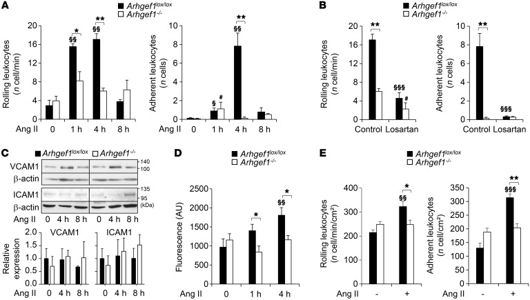
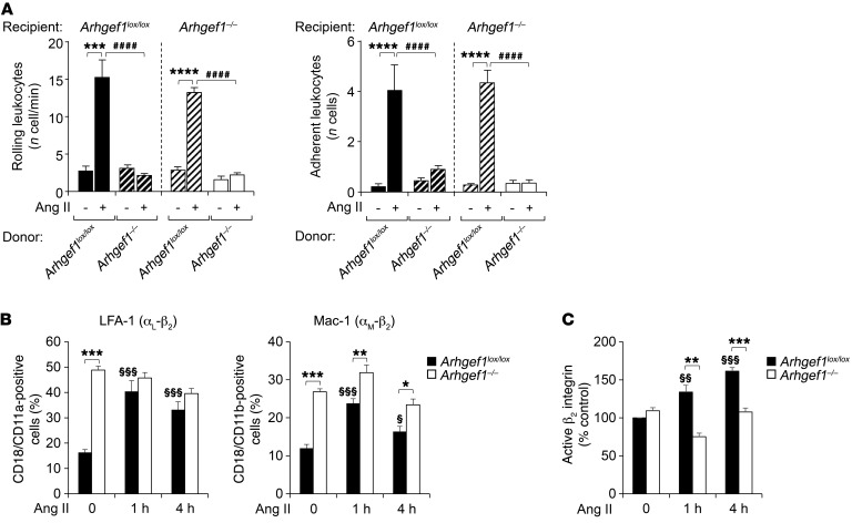
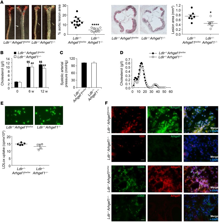
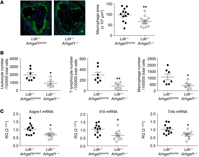
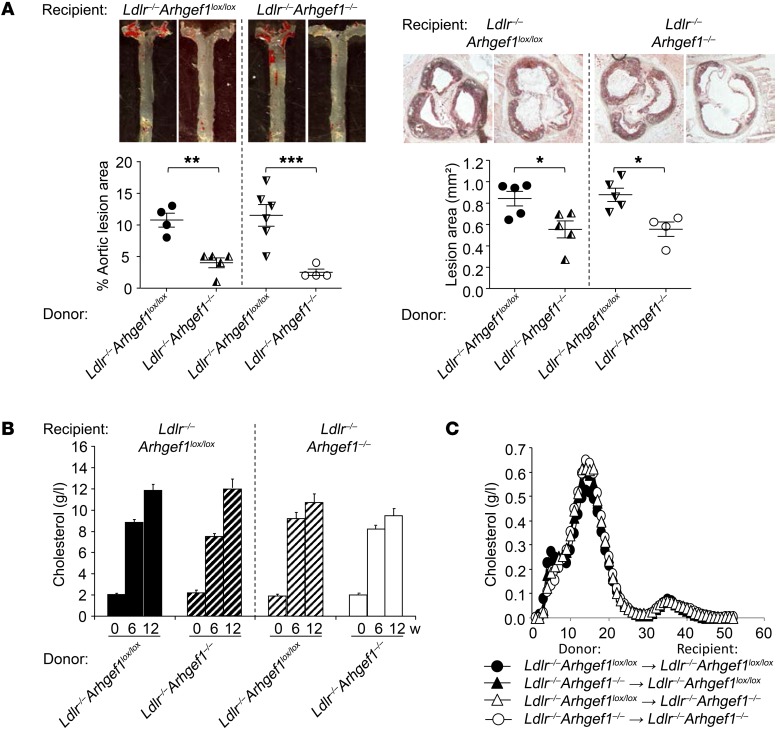
Abnormal activity of the renin-angiotensin-aldosterone system plays a causal role in the development of hypertension, atherosclerosis, and associated cardiovascular events such as myocardial infarction, stroke, and heart failure. As both a vasoconstrictor and a proinflammatory mediator, angiotensin II (Ang II) is considered a potential link between hypertension and atherosclerosis. However, a role for Ang II-induced inflammation in atherosclerosis has not been clearly established, and the molecular mechanisms and intracellular signaling pathways involved are not known. Here, we demonstrated that the RhoA GEF Arhgef1 is essential for Ang II-induced inflammation. Specifically, we showed that deletion of Arhgef1 in a murine model prevents Ang II-induced integrin activation in leukocytes, thereby preventing Ang II-induced recruitment of leukocytes to the endothelium. Mice lacking both LDL receptor (LDLR) and Arhgef1 were protected from high-fat diet-induced atherosclerosis. Moreover, reconstitution of Ldlr-/- mice with Arhgef1-deficient BM prevented high-fat diet-induced atherosclerosis, while reconstitution of Ldlr-/- Arhgef1-/- with WT BM exacerbated atherosclerotic lesion formation, supporting Arhgef1 activation in leukocytes as causal in the development of atherosclerosis. Thus, our data highlight the importance of Arhgef1 in cardiovascular disease and suggest targeting Arhgef1 as a potential therapeutic strategy against atherosclerosis.

Keywords: Atherosclerosis; G-proteins; Integrins; Vascular Biology.


Conflict of interest statement

Conflict of interest: The authors have declared that no conflict of interest exists.

Figures

Figure 1
Figure 1. Deletion of Arhgef1 inhibits leukocyte rolling and adhesion.
(A) Time-dependent in vivo effect of Ang II (30 pmol) on leukocyte rolling and adhesion in mesenteric vessels of Arhgef1lox/lox and Arhgef1–/– mice (n = 5 mice). (B) Effect of losartan on leukocyte rolling and adhesion induced by Ang II (30 pmol, 4 hours) in mesenteric vessels of Arhgef1lox/lox and Arhgef1–/– mice (n = 5 mice). (C) Representative immunoblot of VCAM1, ICAM1, and β-actin in lysates of aortas from Arhgef1lox/lox and Arhgef1–/– mice before (0) and 4 and 8 hours after Ang II treatment (n = 3) and corresponding quantification. All lanes were run on the same gel, but lanes 3 and 4 were noncontiguous as indicated by the black dividing line. (D) In vitro static adhesion of Arhgef1lox/lox and Arhgef1–/– leukocytes on ICAM before (0) and 1 and 4 hours after Ang II treatment (n = 6 experiments). (E) In vitro analysis of Arhgef1lox/lox and Arhgef1–/– leukocyte rolling and adhesion on HUVECs under shear flow, before (–) and 4 hours after (+) Ang II treatment (n = 5). *P < 0.05, **P < 0.01, Arhgef1lox/lox vs. Arhgef1–/– in same condition; §P < 0.05, §§P < 0.01, §§§P < 0.001, relative to the control condition for Arhgef1lox/lox; #P < 0.05, relative to the control condition for Arhgef1–/–; Mann-Whitney.
Figure 2
Figure 2. Deletion of the RhoA exchange factor Arhgef1 in leukocytes inhibits Ang II–induced leukocyte rolling and adhesion, and β2 integrin activation.
(A) In vivo leukocyte rolling and adhesion in chimeric mice before (–) and 4 hours after Ang II treatment (30 pmol) (n = 5). ***P < 0.001, ****P < 0.0001, Ang II vs. control condition for Arhgef1lox/lox BM donor; ####P < 0.0001, Arhgef1lox/lox vs. Arhgef1–/– BM donors in same condition; 1-way ANOVA followed by Sidak post hoc test. (B) Expression of LFA-1 and Mac-1 in leukocytes from Arhgef1lox/lox and Arhgef1–/– mice before (0) and 1 and 4 hours after Ang II treatment (n = 5). (C) Expression of the active, high-affinity β2 integrin in leukocytes from Arhgef1lox/lox and Arhgef1–/– mice before (0) and 1 and 4 hours after Ang II treatment (n = 4). *P < 0.05, **P < 0.01, ***P < 0.001, Arhgef1lox/lox vs. Arhgef1–/– in same condition; §P < 0.05, §§P < 0.01, §§§P < 0.001, relative to the control (0) condition for Arhgef1lox/lox; Mann-Whitney in B and C.
Figure 3
Figure 3. Deletion of the RhoA exchange factor Arhgef1 inhibits atherosclerosis.
(A) Quantification of atherosclerotic lesions in whole aorta and the aortic root (original magnification, ×10) of Ldlr–/–Arhgef1lox/lox and Ldlr–/–Arhgef1–/– mice (*P < 0.05, ****P < 0.0001, Ldlr–/–Arhgef1lox/lox and Ldlr–/–Arhgef1–/– mice). (B) Plasma cholesterol concentration in Ldlr–/–Arhgef1lox/lox and Ldlr–/–Arhgef1–/– mice at the beginning, and after 6 and 12 weeks of high-fat diet (n = 5). §§P < 0.01, relative to the control (0) condition for Arhgef1lox/lox; ##P < 0.01, relative to the control (0) condition for Arhgef1–/–. (C) Systolic blood pressure of Ldlr–/–Arhgef1lox/lox and Ldlr–/–Arhgef1–/– mice after 12 weeks of high-fat diet. (D) FPLC cholesterol profile of Ldlr–/–Arhgef1lox/lox and Ldlr–/–Arhgef1–/– mice after 12 weeks of high-fat diet. (E) Oxidized LDL uptake in peritoneal macrophages from Ldlr–/–Arhgef1lox/lox and Ldlr–/–Arhgef1–/– mice quantified by [3H]-cholesterol uptake and illustrated above by BODIPY staining. (F) Representative images of CD3, CD68, and Arhgef1 staining of aortic root sections from Ldlr–/–Arhgef1lox/lox and Ldlr–/–Arhgef1–/– mice (n = 6; scale bars: 20 μm). Mann-Whitney test in AC and E; 1-way ANOVA followed by Bonferroni post hoc test in D.
Figure 4
Figure 4. Deletion of Arhgef1 decreases immune cell accumulation and inflammation in atherosclerotic aorta.
(A) Representative photomicrographs of CD68 macrophage staining and corresponding quantitative analysis of macrophage accumulation in atherosclerotic lesions of Ldlr–/–Arhgef1lox/lox and Ldlr–/–Arhgef1–/– mice (original magnification, ×10; *P < 0.05, Mann-Whitney test). (B) Quantification of total leukocytes, T lymphocytes, and macrophages in atherosclerotic aorta of Ldlr–/–Arhgef1lox/lox and Ldlr–/–Arhgef1–/– by flow cytometry (*P < 0.05, **P < 0.01, Mann-Whitney test). (C) Measurements by quantitative reverse transcriptase PCR of mRNA levels of the macrophage markers Adgre1, Il1b, and Tnfa (encoding F4/80, IL-1β, and TNF-α, respectively) in atherosclerotic aorta of Ldlr–/–Arhgef1lox/lox and Ldlr–/–Arhgef1–/– (*P < 0.05, Mann-Whitney test). Results are expressed as relative quantity (RQ).
Figure 5
Figure 5. Deletion of the RhoA exchange factor Arhgef1 in leukocytes inhibits atherosclerosis.
(A) Quantification of atherosclerotic lesions in whole aorta and aortic root (original magnification, ×10) of chimeric Ldlr–/– mice (*P < 0.05, **P < 0.01, ***P < 0.001, Ldlr–/–Arhgef1lox/lox donor vs. Ldlr–/–Arhgef1–/– donor in the same recipient Ldlr–/–Arhgef1 genotype). (B) Plasma cholesterol concentration in Ldlr–/– chimeric mice (irradiated Ldlr–/–Arhgef1lox/lox and Ldlr–/–Arhgef1–/– recipient mice transplanted with BM from Ldlr–/–Arhgef1lox/lox and Ldlr–/–Arhgef1–/– donor mice) at the beginning (0), and after 6 and 12 weeks of high-fat diet (n = 5 in each group). (C) FPLC cholesterol profile in Ldlr–/– chimeric mice after 12 weeks of high-fat diet (n = 5 in each group). 1-way ANOVA followed by Bonferroni post hoc test.
Figure 6
Figure 6. Immunostaining of Arhgef1 and immune cells in human carotid artery atherosclerotic lesion.
(A) Immunohistochemical staining showing Arhgef1 and CD3 expression (in brown) in human atherosclerotic carotid artery sections and the correlation of their respective staining area relative to the whole lesion (original magnification, ×2.5). The surrounded area in the complete section is displayed with higher magnification above (×20). (B) Representative coimmunofluorescent staining of Arhgef1 with CD3 (top, T cells), CD20 (middle, B cells), and CD68 (bottom, macrophages), and corresponding plots of Pearson’s correlation coefficients for the colocalization analysis. Colocalization of Arhgef1 and the T cell marker CD3 is illustrated in the higher-magnification image above the graph (arrows). Nuclei are stained with DAPI. Scale bars: 10 μm.

Similar articles

Cited by

References

    1. de Gasparo M, Catt KJ, Inagami T, Wright JW, Unger T. International union of pharmacology. XXIII. The angiotensin II receptors. Pharmacol Rev. 2000;52(3):415–472. - PubMed
    1. Touyz RM, Berry C. Recent advances in angiotensin II signaling. Braz J Med Biol Res. 2002;35(9):1001–1015. doi: 10.1590/S0100-879X2002000900001. - DOI - PubMed
    1. Ferrario CM, Strawn WB. Role of the renin-angiotensin-aldosterone system and proinflammatory mediators in cardiovascular disease. Am J Cardiol. 2006;98(1):121–128. doi: 10.1016/j.amjcard.2006.01.059. - DOI - PubMed
    1. Cassis LA, Rateri DL, Lu H, Daugherty A. Bone marrow transplantation reveals that recipient AT1a receptors are required to initiate angiotensin II-induced atherosclerosis and aneurysms. Arterioscler Thromb Vasc Biol. 2007;27(2):380–386. - PubMed
    1. Daugherty A, Manning MW, Cassis LA. Angiotensin II promotes atherosclerotic lesions and aneurysms in apolipoprotein E-deficient mice. J Clin Invest. 2000;105(11):1605–1612. doi: 10.1172/JCI7818. - DOI - PMC - PubMed

Publication types

MeSH terms