

The Effect of Nutritive and Non-Nutritive Sweeteners on the Growth, Adhesion, and Biofilm Formation of Candida albicans and Candida tropicalis
- PMID: 29131083
- PMCID: PMC5848478
- DOI: 10.1159/000484718
The Effect of Nutritive and Non-Nutritive Sweeteners on the Growth, Adhesion, and Biofilm Formation of Candida albicans and Candida tropicalis
Abstract
Objective: To determine the effect of glucose, sucrose, and saccharin on growth, adhesion, and biofilm formation of Candida albicans and Candida tropicalis.
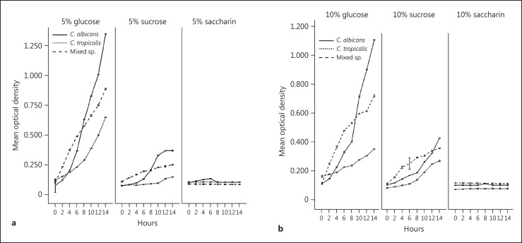
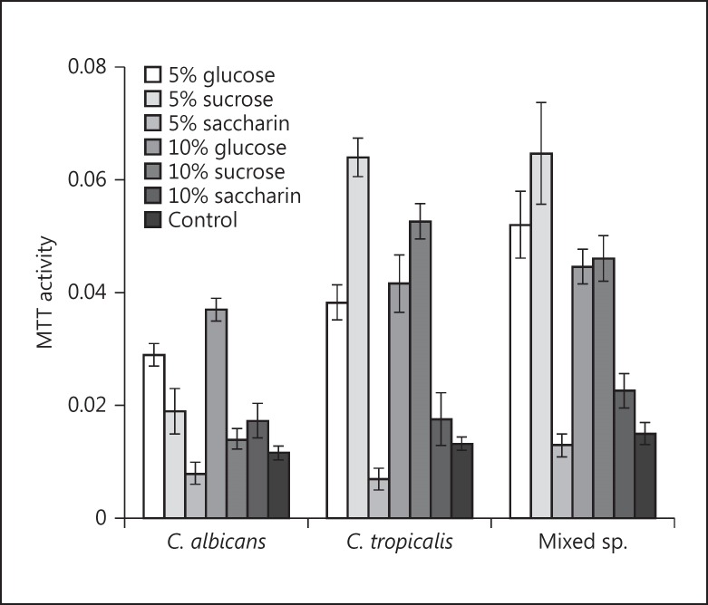
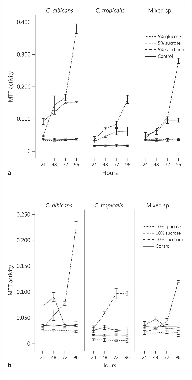
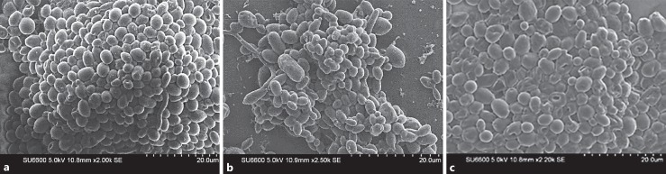
Materials and methods: The growth rates of mono-cultures of planktonic C. albicans and C. tropicalis and 1:1 mixed co-cultures were determined in yeast nitrogen broth supplemented with 5% (30 mM) and 10% (60 mM) glucose, sucrose, and saccharin, using optical density measurements at 2-h intervals over a 14-h period. Adhesion and biofilm growth were performed and the growth quantified using a standard 3-(4,5-dimethylthiazol-2-yl)-2,5-diphenyltetrazolium bromide (MTT) assay. The biofilm architecture was visualized using scanning electron microscopy. One- and two-way analysis of variance (ANOVA) was performed to analyse the differences among multiple means.
Results: The highest planktonic growth was noted in 5% glucose after 14 h (p < 0.05). No significant planktonic growth was observed in either concentration of saccharin. Both the concentrations of glucose and sucrose elicited significantly increased adhesion from MTT activity of 0.017 to >0.019 in mono- as well as co-cultures (p < 0.05), whilst the lower concentration of saccharin significantly dampened the adhesion. Maximal biofilm growth was observed in both species with the lower concentration of sucrose (5%), although a similar concentration of saccharin abrogated biofilm development: the highest MTT value (>0.35) was obtained for glucose and the lowest (>0.15) for saccharin.
Conclusion: In this study, glucose and sucrose accelerated the growth, adhesion, and biofilm formation of Candida species. However, the non-nutritive sweetener saccharin appeared to dampen, and in some instances suppress, these virulent attributes of Candida.
Keywords: Adhesion; Biofilms; Candida species; Glucose; Growth sucrose; Saccharin.
© 2017 The Author(s) Published by S. Karger AG, Basel.
Figures
Similar articles
-
Culture media profoundly affect Candida albicans and Candida tropicalis growth, adhesion and biofilm development.Mem Inst Oswaldo Cruz. 2016 Nov;111(11):697-702. doi: 10.1590/0074-02760160294. Epub 2016 Oct 3. Mem Inst Oswaldo Cruz. 2016. PMID: 27706381 Free PMC article.
-
Thymus vulgaris essential oil and thymol inhibit biofilms and interact synergistically with antifungal drugs against drug resistant strains of Candida albicans and Candida tropicalis.J Mycol Med. 2020 Apr;30(1):100911. doi: 10.1016/j.mycmed.2019.100911. Epub 2019 Nov 7. J Mycol Med. 2020. PMID: 32008964
-
Interactions between Terpinen-4-ol and Nystatin on biofilm of Candida albicans and Candida tropicalis.Braz Dent J. 2018 Jul-Aug;29(4):359-367. doi: 10.1590/0103-6440201802073. Braz Dent J. 2018. PMID: 30462762
-
Adhesion and biofilm formation by the opportunistic pathogen Candida tropicalis: what do we know?Can J Microbiol. 2023 Jun 1;69(6):207-218. doi: 10.1139/cjm-2022-0195. Epub 2023 Feb 21. Can J Microbiol. 2023. PMID: 36809069 Review.
-
Alteration of oral microbial biofilms by sweeteners.Biofilm. 2023 Dec 13;7:100171. doi: 10.1016/j.bioflm.2023.100171. eCollection 2024 Jun. Biofilm. 2023. PMID: 38197082 Free PMC article. Review.
Cited by
-
Amperometric microbial biosensor for sugars and sweetener classification using principal component analysis in beverages.J Food Sci Technol. 2023 Jan;60(1):382-392. doi: 10.1007/s13197-022-05625-8. Epub 2022 Nov 24. J Food Sci Technol. 2023. PMID: 36618051 Free PMC article.
-
Antifungal Effect of Poly(methyl methacrylate) Coated with Polyelectrolyte Multilayers.ACS Omega. 2025 May 7;10(19):19832-19839. doi: 10.1021/acsomega.5c00883. eCollection 2025 May 20. ACS Omega. 2025. PMID: 40415842 Free PMC article.
-
Interactions of Non-Nutritive Artificial Sweeteners with the Microbiome in Metabolic Syndrome.Immunometabolism. 2022;4(2):e220012. doi: 10.20900/immunometab20220012. Epub 2022 Apr 18. Immunometabolism. 2022. PMID: 35528135 Free PMC article.
-
Influence of Laboratory Culture Media on in vitro Growth, Adhesion, and Biofilm Formation of Pseudomonas aeruginosa and Staphylococcus aureus.Med Princ Pract. 2019;28(1):28-35. doi: 10.1159/000494757. Epub 2018 Oct 23. Med Princ Pract. 2019. PMID: 30352435 Free PMC article.
-
Exploring the Potential of Candida sp. SW4-6 as a Probiotic for Enhancing Water Quality in Aquaculture.Microorganisms. 2024 Dec 29;13(1):42. doi: 10.3390/microorganisms13010042. Microorganisms. 2024. PMID: 39858810 Free PMC article.
References
-
- Samaranayake LP. Host factors and oral candidosis. In: Samaranayake LP, MacFarlane TW, editors. Oral Candidosis. London: Butterworth; 1990. pp. 66–103.
-
- Sardi JC, Scorzoni L, Bernardi T, et al. Candida species: current epidemiology, pathogenicity, biofilm formation, natural antifungal products and new therapeutic options. J Med Microbiol. 2013;62:10–24. - PubMed
-
- Weerasekera MM, Sissons CH, Wong L, et al. Use of denaturing gradient gel electrophoresis for the identification of mixed oral yeasts in human saliva. J Med Microbiol. 2013;62:319–330. - PubMed
MeSH terms
Substances
LinkOut - more resources
Full Text Sources
Other Literature Sources
