. 2018 Jan;14(1):42-49.
doi: 10.1038/nchembio.2498. Epub 2017 Nov 13.

Nonimmune cells equipped with T-cell-receptor-like signaling for cancer cell ablation

Affiliations

Nonimmune cells equipped with T-cell-receptor-like signaling for cancer cell ablation

Ryosuke Kojima et al. Nat Chem Biol. 2018 Jan.

Abstract

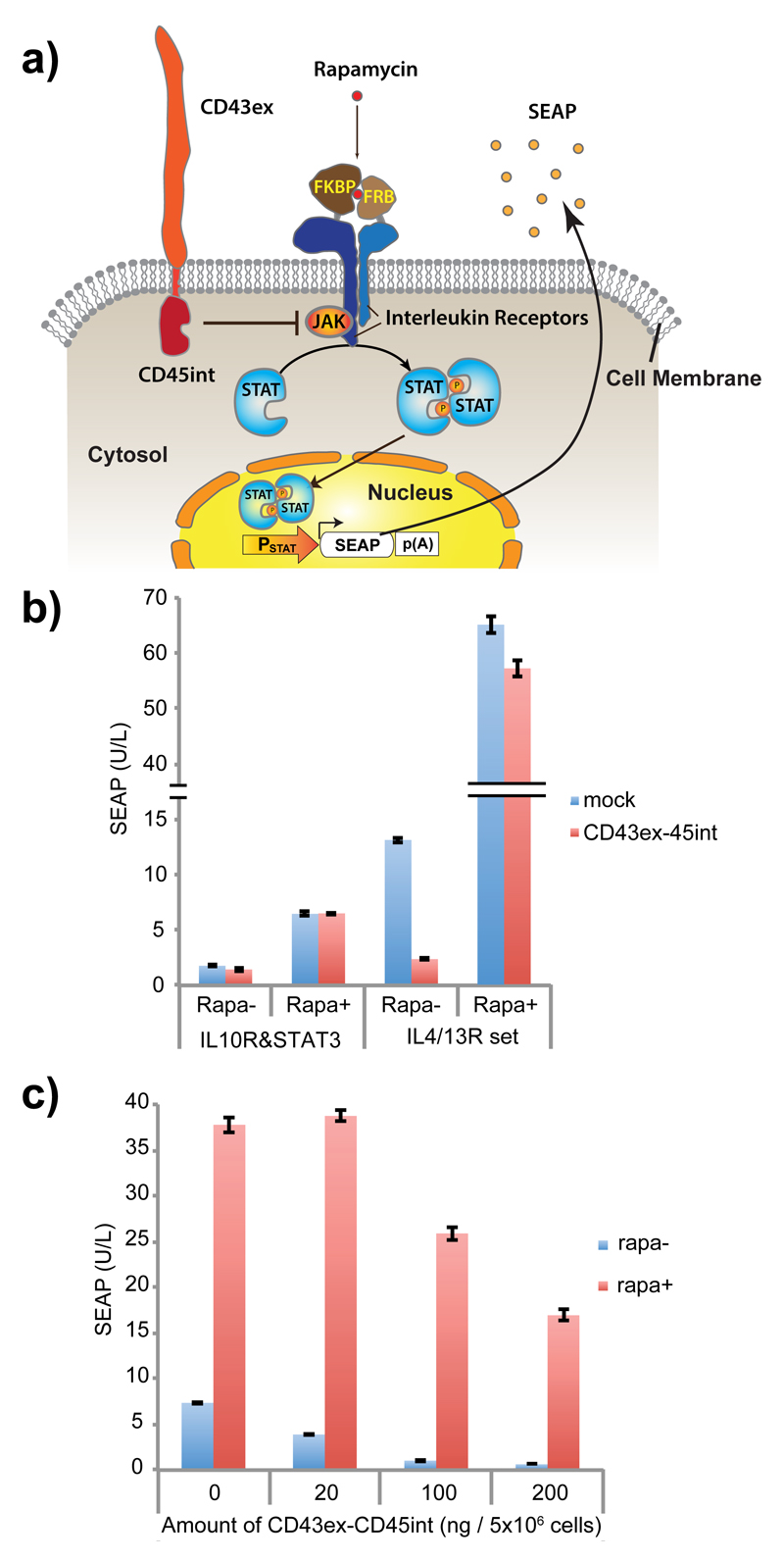
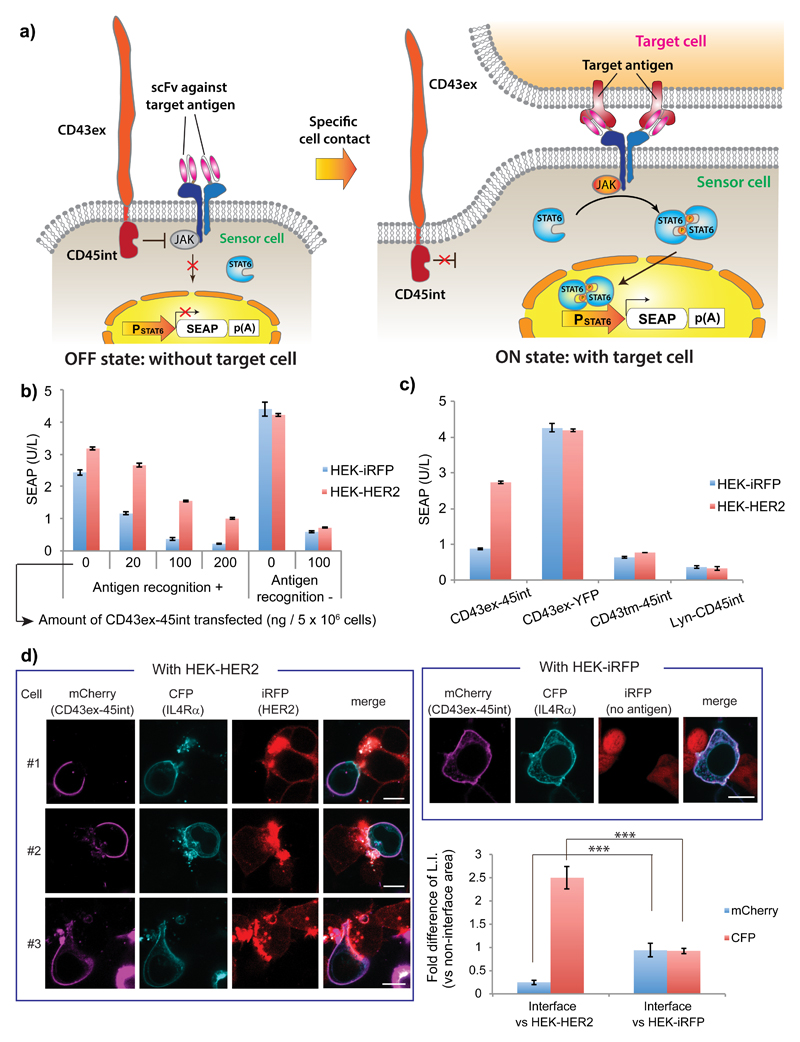
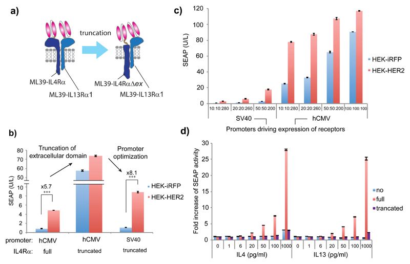
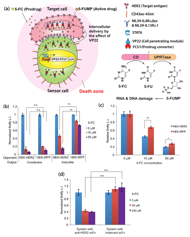
The ability to engineer custom cell-contact-sensing output devices into human nonimmune cells would be useful for extending the applicability of cell-based cancer therapies and for avoiding risks associated with engineered immune cells. Here we have developed a new class of synthetic T-cell receptor-like signal-transduction device that functions efficiently in human nonimmune cells and triggers release of output molecules specifically upon sensing contact with a target cell. This device employs an interleukin signaling cascade, whose OFF/ON switching is controlled by biophysical segregation of a transmembrane signal-inhibitory protein from the sensor cell-target cell interface. We further show that designer nonimmune cells equipped with this device driving expression of a membrane-penetrator/prodrug-activating enzyme construct could specifically kill target cells in the presence of the prodrug, indicating its potential usefulness for target-cell-specific, cell-based enzyme-prodrug cancer therapy. Our study also contributes to the advancement of synthetic biology by extending available design principles to transmit extracellular information to cells.


Conflict of interest statement

Competing financial interests

The authors declare no competing financial interests.

Figures

Figure 1
Figure 1. Evaluation of CD43ex-45int for suppressing cytokine receptor-mediated signaling pathways.
(a) Schematic illustration of the design. In the presence of rapamycin (rapa), Janus kinase (JAK) is activated by receptor dimerization, leading to phosphorylation and dimerization of signal transducer and activator of transcription (STAT). Dimerized STAT translocates to the nucleus, and promotes transgene expression via a STAT-responsive minimal promoter. The effect of co-expression of CD43ex-45int on signaling was determined by quantifying induced expression of a reporter protein, secreted alkaline phosphatase (SEAP). (b) SEAP expression from HEK-293T cells co-transfected with pRK96 (PhCMV-CD43ex-45int-pA) (pA: poly adenylation signal) (or pcDNA3.1(+) for mock) and interleukin receptors together with corresponding STAT and its reporter (IL10R & STAT3 set: pLeo56 (PhCMV-FKBP-IL10Rα-pA), pLeo57 (PhCMV-FRB-IL10Rβ-pA), pLS15 (PhCMV-STAT3-pA) and pLS13 (PSTAT3-SEAP-pA). IL4/13R & STAT6 set: pLeo53 (PhCMV-FKBP-IL4Rα-pA) and pLeo52 (PhCMV-FRB-IL13Rα1-pA), pLS16 (PhCMV-STAT6-pA), and pLS12 (PSTAT6-SEAP-pA)) (± rapa as inducer). (c) Dose-dependency of the inhibitory activity of CD43ex-45int on IL4/13R signaling. SEAP expression with different amounts of pRK96 is shown. All the data are mean ± SEM of three independent experiments measured in triplicate (n=3).
Figure 2
Figure 2. Development of the specific-cell-contact-sensing device.
(a) Schematic illustration. Without target cells, JAK is inhibited by CD43ex-CD45int and downstream signaling is shut off. When the sensor cell binds to a target cell via scFv-antigen interaction, CD43ex-45int is segregated from the cell-cell interface, which turns on downstream signaling (translocation of phosphorylated STAT6 dimer to the nucleus, and STAT-6-responsive transgene expression). (b) Evaluation of effect of antigen recognition. Sensor HEK-293T cells were transfected with IL4Rα and IL13Rα1 bearing, or not bearing an antigen recognition moiety, together with various amounts of pRK96 (see Table 1). After mixing them with HEK-HER2 or HEK-iRFP cells, SEAP secreted from the sensor cells was assayed. (c) Assays to verify CD43ex-45int function. Sensor HEK-293T cells were transfected with pRK96, pRK14 (PhCMV-CD43ex-YFP-pA), pRK290 (PhCMV-CD43tm-45int-pA), or pRK291 (PhCMV-Lyn-CD45int-pA) as well as the other signaling devices (see Table 1). Sensor cells were mixed with HEK-HER2 or HEK-iRFP cells, and SEAP secreted from the sensor cells was assayed. Data in Fig. 2b,c are mean ± SEM of three independent experiments measured in triplicate (n=3). (d) Imaging analysis of CD43ex-45int segregation (scale bar: 10 µm). Sensor HEK-293T cells were transfected with pRK293 (PhCMV-ML39-IL10RαΔint-CFP-pA) and pHCM-CD43ex-CD45int-mCherry (LTR-PhCMV-CD43ex-45int-mCherry-LTR), and mixed with HEK-HER2 or HEK-iRFP cells. Statistical analysis of localization of CD43ex-45int-mCherry and ML39-IL10RαΔint-CFP was also conducted. Data represent average fluorescence intensity of each fluorophore at the cell-cell interface (normalized to the intensity at regions of the plasma membrane without cell contacts) ± SEM of 9 different cells. ***P<0.001 (n=9), two-tailed Student’s t-test.
Figure 3
Figure 3. The optimized specific-cell-contact-sensing system.
(a) Schematic illustration of receptor truncation. The receptor set bearing truncated IL4R (IL4RαΔex) does not need ligands for activation. Therefore, output gene expression is increased, and the system is non-responsive to native ligands. (b) Evaluation of the effect of receptor truncation and promoter optimization. The sensor HEK-293T cells were transfected as shown in Table 1. After mixing them with HEK-HER2 or HEK-iRFP cells, SEAP secreted from the sensor cells was assayed. (c) Effect of dose dependency of each component on the system performance. Different amounts of plasmids encoding receptors (driven by either PSV40 or PhCMV) and CD43ex-45int were transfected in sensor HEK-293T cells (See Table 1). Numbers under each column (10:10:280 etc) indicate the amounts (ng/5x105 cells) of transfected plasmids encoding IL4RαΔex (pRK122 or pRK119), IL13Rα1 (pRK123 or pRK114), and CD43ex-45int (pRK96), respectively from left to right. After mixing the sensor cells with HEK-HER2 or HEK-iRFP cells, SEAP secreted from the sensor cells was assayed. (d) System responsiveness to native ligands (IL4, IL13) with different receptor sets. The sensor HEK-293T cells were transfected as shown in Table 1b. SEAP secreted from the sensor cells in the presence of various concentrations of IL4 and IL13 was assayed. All the data are mean ± SEM of three independent experiments measured in triplicate (n=3). Two-tailed Student’s t-tests were conducted for Fig.3b. ***P<0.001.
Figure 4
Figure 4. Application to target-cell-specific activatable enzyme-prodrug cancer therapy.
(a) Schematic illustration. Sensor cells express effector protein VP22-FCU1, which is delivered to adjacent cells by VP22 and converts prodrug 5-FC into cytotoxic 5-FUMP via its cytosine deaminase (CD) and uracil phosphoribosyltransferase (UPRTase) activities. This occurs only when sensor cells contact target cells expressing specific antigen. (b) Demonstration of target-specific cell killing. Sensor HEK-293T cells were transfected with pRK96, pRK122 (PSV40-ML39-IL4RαΔex-pA), pRK123 (PSV40-ML39-IL13Rα1-pA), and pLS16, plus either pRK131 (PhCMV-VP22-FCU1-pA) (“constitutive”) or pRK223 (PSTAT6-VP22-FCU1-pA) (“inducible”). Transfected sensor cells were mixed with HEK-HER2-Luc or HEK-iRFP-Luc cells in 3D culture. 5-FC (0-50 µM) was added. After 4 days, viability was measured as luminescence intensity (normalized to no 5-FC condition). (c) Target-specific cell killing in mixed culture. Sensor HEK-293T cells were transfected as in Fig. 4b (inducible), then mixed with 1:1 HEK-HER2-Luc and HEK-iRFP (for measuring HEK-HER2-Luc viability), or 1:1 HEK-HER2 and HEK-iRFP-Luc (for measuring HEK-iRFP-Luc viability) in 3D culture. 5-FC (0-50 µM) was added, and viability was measured as above. (d) SKBR3 cell killing by hMSC-TERT cells equipped with specific-cell-contact-sensing system. hMSC-TERT cells were transfected with the same components as in Fig. 4b (inducible) (system with anti-HER2), or components having irrelevant scFv (pRK145 (PSV40-SP6-IL4RαΔex-pA) and pRK144 (PSV40-SP6-IL13Rα1-pA)), then mixed with SKBR3 cells stably expressing firefly luciferase. After addition of 5-FC (0-250 µM), the same assay as in (b) was conducted. Data are mean ± SEM of three independent experiments measured in triplicate (n=3). Two-tailed Student’s t-test: *P<0.05, **P<0.01, ***P<0.001. n.s: P>0.05.

Comment in

Similar articles

Cited by

References

    1. Almasbak H, Aarvak T, Vemuri MC. CAR T Cell Therapy: A game changer in cancer treatment. J Immunol Res. 2016;2016:5474602. - PMC - PubMed
    1. Jackson HJ, Rafiq S, Brentjens RJ. Driving CAR T-cells forward. Nat Rev Clin Oncol. 2016;13:370–383. - PMC - PubMed
    1. June CH, Blazar BR, Riley JL. Engineering lymphocyte subsets: tools, trials and tribulations. Nat Rev Immunol. 2009;9:704–716. - PMC - PubMed
    1. Kalaitsidou M, Kueberuwa G, Schutt A, Gilham DE. CAR T-cell therapy: toxicity and the relevance of preclinical models. Immunotherapy. 2015;7:487–497. - PubMed
    1. Kojima R, Aubel D, Fussenegger M. Novel theranostic agents for next-generation personalized medicine: small molecules, nanoparticles, and engineered mammalian cells. Curr Opin Chem Bio l. 2015;28:29–38. - PubMed

Substances