. 2018 Jan;47(2):140-149.
doi: 10.1111/ejn.13778.

Interferon regulatory factor 4/5 signaling impacts on microglial activation after ischemic stroke in mice

Affiliations

Interferon regulatory factor 4/5 signaling impacts on microglial activation after ischemic stroke in mice

Abdullah Al Mamun et al. Eur J Neurosci. 2018 Jan.

Abstract

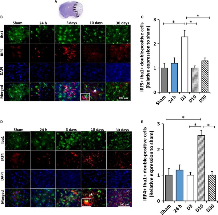
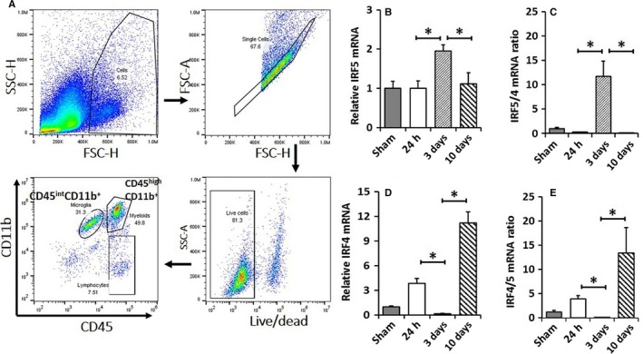
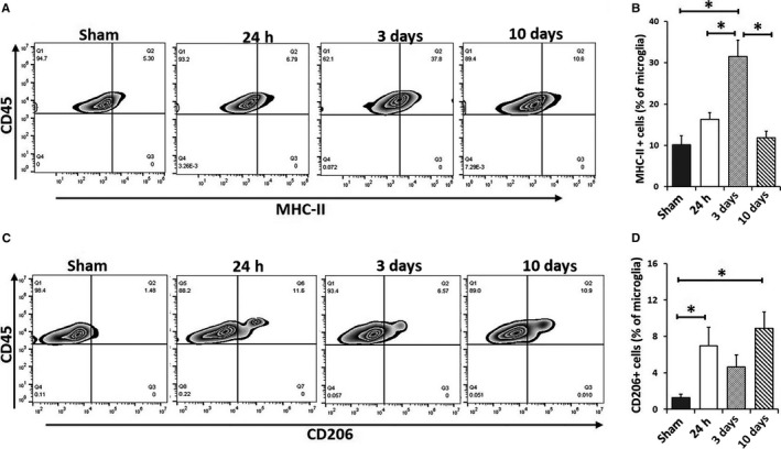
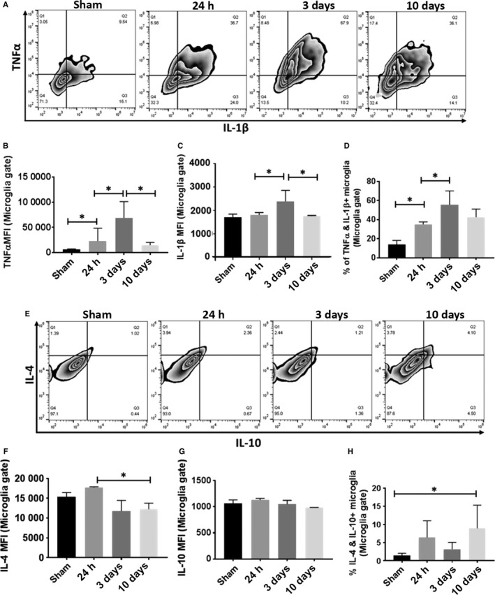
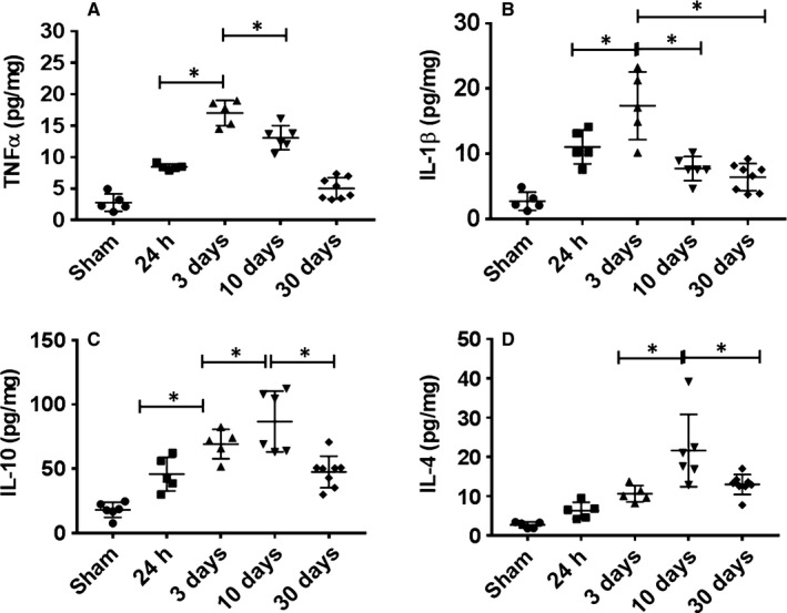
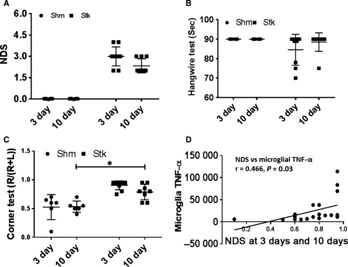
Microglial activation is a key element in initiating and perpetuating inflammatory responses to stroke. Interferon regulatory factor 5 (IRF5) and IRF4 signaling have been found critical in mediating macrophage pro-inflammatory (M1) and anti-inflammatory (M2) phenotypes, respectively, in peripheral inflammation. We hypothesize that the IRF5/4 regulatory axis also mediates microglial activation after stroke. C57BL6 mice of 8-12 weeks were subject to a 90-min middle cerebral artery occlusion, and the brains evaluated at 24 h, 3, 10 and 30 days after reperfusion. Flow cytometry was utilized to examine microglial activation and cytokine expression. RT-PCR was performed for mRNA levels of IRF5/4 in sorted microglia. Microglial expression of IRF5/4 was examined by immunohistochemistry, and brain cytokine levels were determined by ELISA. Our results revealed that the IRF5 mRNA level in sorted microglia increased at 3 days of stroke; whereas IRF4 mRNA level exhibited biphasic increases, with a transient rise at 24 h and a peak at 10 days. The same pattern was seen in IRF5/4 protein colocalization with Iba-1+ cells by IHC. Intracellular levels of TNF-α and IL-1β in microglia peaked at 3 days of stroke, and IL-4+ IL-10+ double-positive microglia significantly increased at day 10. Brain levels of these cytokines were consistent with microglial cytokine changes. Worse behavior test results were seen at 3 days vs. 10 days of stroke. We conclude that microglia phenotypes are dynamic to ischemic stroke, and IRF5/4 signaling may regulate microglial M1/M2 activation and impact on stroke outcomes.

Keywords: IRF4; IRF5; ischemic stroke; microglia; neuroinflammation.


Figures

Figure 1
Figure 1
IRF5/4 expression in activated microglia/macrophages at 24 h, 3, 10 and 30 days following 90‐min MCAO. Image analysis was performed in eight ipsilateral regions (black boxes) at the inner boundary zone of the infarct (A). Co‐expression of IRF5 or IRF4 (red) with Iba1 (green) in activated microglia/macrophages was shown in Figure (B) and (D), respectively. IRF5+Iba1+ and IRF4+Iba1+ (inset in the d10 merged) double‐positive cells in the peri‐infarct area of the ipsilateral hemisphere of stroke mice were quantified and showed in (C) and (E), respectively. White arrowheads indicate the double‐positive cell as shown in the inset box. All the quantification data were presented as relative changes compared to sham as fold change. = 6 stroke/4 sham animals per group; *< 0.05. [Colour figure can be viewed at http://wileyonlinelibrary.com].
Figure 2
Figure 2
IRF5/4 mRNA levels in activated microglia. A representative dot plot depicts the gating strategy used to sort microglia (A). CD45int CD11b+ cells were considered as microglia. Results of Q‐PCR for mRNA expression were shown in (B) for IRF5 and in (D) for IRF4. mRNA ratios of IRF5/IRF4 and IRF4/IRF5 were shown in (C) and (E), respectively. = 6 stroke/4 sham animals per group; *< 0.05. [Colour figure can be viewed at http://wileyonlinelibrary.com].
Figure 3
Figure 3
Microglia activation after 90‐min MCAO. Quantification of MHCII + microglia (A, B) and CD206+ microglia percentage (C, D) of total gated microglia at 24 h, 3 and 10 days after MCAO. = 6 stroke/4 sham animals per group; *< 0.05.
Figure 4
Figure 4
Measurement of intracellular pro‐ and anti‐inflammatory cytokines in microglia at 24 h, 3 and 10 days following 90‐min MCAO. Representative zebra plots (A) and (E) showed microglial production of pro‐inflammatory TNF‐α/IL‐1β and anti‐inflammatory IL‐4/IL‐10, respectively. The expression levels of TNF‐α, IL‐1β, IL‐4 and IL‐10 were measured as mean fluorescence intensity (MFI) in (B), (C), (F) and (G), respectively. Quantification of TNF‐α+ IL‐1β+ and IL‐4+ IL‐10+ double‐positive microglia was showed in (D) and (H), respectively. = 8 stroke/5 sham animals per group; *< 0.05.
Figure 5
Figure 5
Cytokine levels in the ischemic brain. Pro‐inflammatory (TNFα and IL‐1β) (A and B) and anti‐inflammatory (IL‐10 and IL‐4) (C and D) cytokine levels (pg/mg of protein) were measured in the ipsilateral hemispheres of sham and stroke mice at 24 h, 3, 10 and 30 days after reperfusion. = 6 stroke/4 sham animals per group; *< 0.05.
Figure 6
Figure 6
Assessment of behavior outcomes at 3 and 10 days following 90‐min MCAO. Neurological deficit score (NDS) (A), hangwire (B) and corner test (C) were assessed for each group at 3 and 10 days. Microglial intracellular TNF‐α significantly correlated with at 3 and 10 days after stroke (D). = 6–8 animals per group; *< 0.05.

References

    1. Al Mamun, A. & Liu, F. (2017) Role of IRF4‐mediated inflammation: implication in neurodegenerative diseases. Neurol. Neurotherapy Open Access J., 2, 000107. - PMC - PubMed
    1. Balkhi, M.Y. , Fitzgerald, K.A. & Pitha, P.M. (2010) IKKalpha negatively regulates IRF‐5 function in a MyD88‐TRAF6 pathway. Cell. Signal., 22, 117–127. - PubMed
    1. Burke, N.N. , Kerr, D.M. , Moriarty, O. , Finn, D.P. & Roche, M. (2014) Minocycline modulates neuropathic pain behaviour and cortical M1‐M2 microglial gene expression in a rat model of depression. Brain Behav. Immun., 42, 147–156. - PubMed
    1. Chen, Y. , Won, S.J. , Xu, Y. & Swanson, R.A. (2014) Targeting microglial activation in stroke therapy: pharmacological tools and gender effects. Curr. Med. Chem., 21, 2146–2155. - PMC - PubMed
    1. Cherry, J.D. , Olschowka, J.A. & O'Banion, M.K. (2014) Neuroinflammation and M2 microglia: the good, the bad, and the inflamed. J. Neuroinflammation, 11, 98. - PMC - PubMed

Publication types

LinkOut - more resources